Veins
With some notable exceptions, veins accompany arteries of the same name. These “satellite” veins are always larger than their respective arteries and frequently duplicated. For example, the brachial artery carrying blood to the forearm and digit may be accompanied by two or more brachial veins returning the blood to the heart.
Some veins are superficial, visible in the subcutaneous tissues, and these are particularly of interest as they may be accessed via venipuncture (introducing a needle into a vein). As indicated earlier, nearly all systemic veins eventually drain into either the cranial vena cava or caudal vena cava.Cranial Vena Cava
The cranial vena cava drains the head, neck, thoracic limbs, and part of the thorax. Tributaries to the cranial vena cava include the jugular veins (internal and external), subclavian veins, and vertebral veins. The external jugular veins
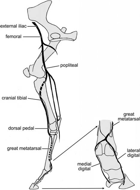
Figure 17-9. Arteries of the pelvic limb in the horse. Primary arterial supply to the pes of the horse is the dorsal metatarsal artery III.
drain the face and much of the head, while the internal jugular veins, if present, along with the vertebral veins drain most of the blood from the brain. Each subclavian vein receives venous blood from the same areas that are supplied by the subclavian artery and its branches (shoulder, neck, and thoracic limbs). The azygos vein (the word azygos derives from the Greek word meaning “unpaired”) lies adjacent to the vertebral column, receiving the segmentally arranged intercostal veins. In horses, the right azygos vein empties at the junction between cranial vena cava and right atrium. Ruminants sometimes have both right and left azygos veins, but more usually have a single left azygos vein, which empties directly into the right atrium with the coronary sinus.
The pig possesses a left azygous vein, which empties into the coronary sinus.Caudal Vena Cava
The caudal vena cava is formed in the abdomen by the junction of the paired internal and external iliac veins. These drain the gluteal and perineal regions and the pelvic limbs, respectively. The caudal vena cava also receives lumbar veins, testicular or ovarian veins, renal veins, and various others from structures associated with the body walls. Just caudal to the point at which the caudal vena cava passes through the caval foramen of the diaphragm, it receives a number of short hepatic veins directly from the liver.
Portal System
A portal system is one in which a vessel divides into capillaries, recombines to form another vessel, and then redivides into a second capillary bed. The hypothalamohypophysial portal system was described in Chapter 12 in relation to the pituitary gland. in birds and in some reptiles and amphibians, part of the venous blood returning from the pelvic limbs enters the kidneys to form a renal portal system (see Chapter 30).
In the hepatic portal system, blood that has perfused the capillary beds of the viscera is brought to the liver by a single large vein, the portal vein, and then is redistributed into a second capillary bed within the substance of the liver (Fig. 17-5).
Tributaries to the portal vein include the gastric vein from the stomach, the splenic vein from the spleen, the mesenteric veins from the intestines, and the pancreatic veins from the pancreas. The portal vein enters the liver and immediately breaks up into smaller and smaller branches there, finally ending in the sinusoids of the liver. Here the blood comes into direct contact with cells of the liver. After being acted upon by the liver cells (see Chapter 21), the blood passes from the sinusoids of the liver into the liver’s venous system and eventually empties into the caudal vena cava.