THE ABOMASUM
The abomasum lies flexed on the abdominal floor, embracing the lower pole of the omasum from behind (Figure 28-7/3). The larger of the two limbs forms a piriform sac that reaches forward to the left to make contact with the body wall between the reticulum and the atrium and ventral sac of the rumen (Figure 28-4, A/6).
This limb is divided by analogy with the simple stomach into fundus and body, but the boundary between these parts is imprecise. In fact, the location of the omasoabomasal opening in the living animal is not known with certainty; it is possible that it is terminal, and in that case no blind diverticulum and therefore no true fundus exists. The cranial part of the fundus is extensively connected to the reticulum, atrium, and ventral sac by muscle bundles.The narrower and more uniform distal limb constitutes the pyloric part of the organ. It passes transversely, or with a slightly cranial inclination, toward the right body wall and ascends to terminate at the pylorus, caudal to the lower part of the omasum (Figure 28-4, D/15). The abomasum does not usually come into contact with the liver in adult cattle.
The abomasum of the sheep and the goat is relatively large. In contrast to the situation in adult cattle, it is usually allowed direct contact with the liver by the smaller size of the omasum.
The position and relations of the abomasum depend on the fullness of the different parts of the stomach, intrinsic abomasal activity, and, most importantly, the contractions of the rumen and reticulum to which the abomasum is attached. Age and pregnancy are also an influence (Figure 28-20). Although it is difficult to specify abomasal relations exactly, it is vital to appreciate that there are limits beyond which deviations produce digestive disturbance and may endanger life. Abomasal displacement, which may be to the right or left, is a well-recognized disorder, particularly in dairy cows (see further on).
The abomasum is lined by a pink, slime-covered, glandular mucosa that is in striking contrast to the harsh lining of the forestomach. At the omasoabomasal junction the epithelium changes abruptly to a simple colum-
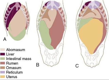
Figure 28-20 Ventral views of the abdominal viscera of a newborn calf (A), a 5-year-old cow (B), and a 6-year-old heavily pregnant cow (C) based on reconstructions of transverse sections of animals frozen in the standing position.
nar epithelium with occasional goblet cells. The lamina propria is less dense than that of the omasum, and frequently, solitary lymph nodules are observed at the junction with the epithelium. The mucosa of the abomasum has all the characteristics of that of the simple stomach (Figure 28-21, A-C). The area is increased about sixfold by the presence of almost a dozen large folds that arise around the entrance and course over the walls of the fundus and body before subsiding as the flexure is approached (Figure 28-22/2). Approximation of the proximal ends of these folds forms a mucosal valve or “plug” that discourages the reflux of ingesta into the omasum. The mucosa of the pyloric part is most remarkable for the large swelling or torus that projects from the lesser curvature to narrow the pyloric passage (Figure 28-22/6). The vascular arrangements within the torus suggest that it is capable of a form of erection, but the possible functional significance of this (and of the entire structure, for that matter) is unknown. The dark mucosa of the body and fundus contains true peptic glands; the glands of the lighter pyloric part secrete mucus alone.
The abomasal wall is relatively thin. The serous covering is deficient only at the attachment to the other stomach chambers and along the origins of the omenta. The muscle coat consists of longitudinal and circular strata. The longitudinal muscle is confined to the curvatures of the fundus and body but forms a thicker and wider covering for the pyloric part.
The circular fibers provide a more complete layer that is better developed over the pyloric part, especially distally.The movements of the adult abomasum are rather sluggish. They consist of general contractions of the proximal limb and more forceful peristalsis confined to the pyloric part. The latter activity often appears to be prompted by the tipping of the ingesta toward the pylorus when the fundic region is elevated by reticular contraction. It is possible that these normal alterations in position facilitate morbid displacements. Atony, with the accumulation of gas in the fundus, is a constant finding in these cases, and it may be that a slight initial displacement is worsened because this gas is denied its usual escape through the omasoabomasal opening when this comes to lie below the gas bubble.
Displacements are commonly related to the high proportion of concentrates to roughage in the ration of stabled cows, which leads to atony of the abomasum and accumulation of liquid ingesta and gas. Pregnancy may be a predisposing factor (Figure 28-20, C). Because the abomasum is well fixed proximally to the heavy omasum and distally by the lesser omentum, it is its middle part that travels farthest from its usual position on the abdominal floor. Contractions of the ruminore- ticulum may allow the abomasum, buoyed by the gas within, to work its way under the atrium of the rumen
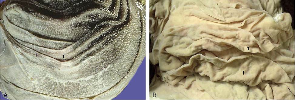
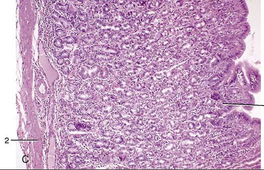
Figure 28-21 A, Internal surface of omasum (cow). 1, Omasal laminae. B, Internal surface of abomasum (cow). 1, Abomasal folds. C, Abomasum (goat) (70?). 1, Gastric pit; 2, lamina muscularis mucosae.
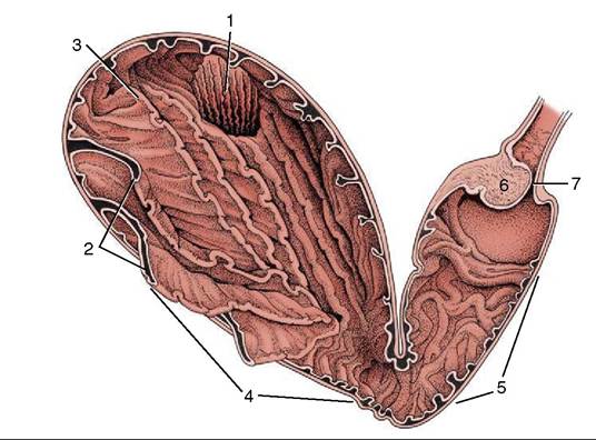
Figure 28-22 Opened abomasum as seen from behind, above, and slightly from the left. 1, Omasoabomasal opening through which the omasal laminae can be seen; 2, abomasal folds; 3, fundus; 4, body; 5, pyloric part; 6, torus pyloricus; 7, pylorus.
and up on the left side. The loop formed by the middle part of the abomasum eventually comes to lie between the rumen and the left abdominal wall, deep to the last three or four ribs, where it can be identified by simultaneous percussion and auscultation (left displacement of the abomasum; LDA). In right displacement (RDA) the loop formed by the middle part of the abomasum slides to the right and lies between the right abdominal wall and the intestines and liver. Displacements to the right are often complicated by twisting of the loop. Treatment of uncomplicated displacements consists of returning the abomasum to its normal position by placing the cow on her back, by deflating the organ through a paramedian incision of the abdominal wall, and by including its muscular coat in the closing of the incision (abomasopexy).