THE OMENTA
The attachment of the greater omentum begins dorsal to the esophagus. The two serosal sheets of which it is composed pass directly onto the rumen but are so
widely separated that the immediately postcardiac part of the rumen roof is enabled to attach directly to the abdominal roof (Figure 28-13/72).
This retroperitoneal space is closed caudally where the two serosal sheets come together halfway along the right longitudinal groove to form a conventional duplicature attaching to the stomach. The attachment of this fold may be traced along the right longitudinal groove, through the caudal groove between the caudal blind sacs, and then forward along the left longitudinal groove. It now crosses the atrium ruminis and widens to make a broad attachment to the reticulum before bending sharply to the right, ventral to the ruminoreticulum, to reach the greater curvature of the abomasum (Figures 28-4, A,C and 28-8/8). It follows this to the pylorus and continues onto the caudal aspect of the first (vertical) part of the duodenum from which it extends onto the descending duodenum and later the mesoduodenum. The omental attachment is reflected where the duodenum turns cra- nially, and it retraces its attachment along the descending duodenum until carried back to the cranial duodenal flexure at the porta of the liver. It then returns to the right face of the rumen via the pancreas.The lesser omentum arises from the visceral surface of the liver, between the porta and the esophageal impression (Figure 28-23), and passes to the region of the reticular groove, the right face of the omasum, and thence along the lesser curvature of the abomasum to
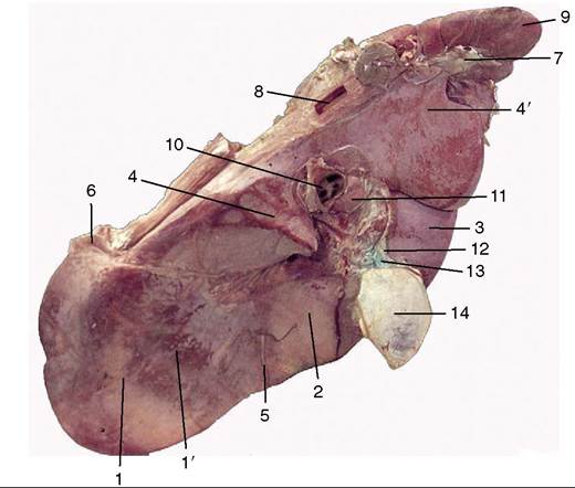
Figure 28-23 Visceral surface of the bovine liver. 1, Left lobe; 1', omasal impression; 2, quadrate lobe; 3, right lobe; 4, 4', papillary and caudate processes of caudate lobe; 5, round ligament; 6, left triangular ligament; 7, right triangular ligament; 8, caudal vena cava; 9, right kidney; 10, portal vein; 11, hepatic lymph node; 12, bile duct; 13, cystic duct; 14, gallbladder.
the first part of the duodenum, which returns it to the liver (Figure 28-4, C).
The omental sheets enclose a space, the omental bursa, which is completely divided from the greater peritoneal cavity except at the epiploic foramen near the porta of the liver. The bursa is a mere capillary cleft in life, but it is simpler for descriptive purposes to envisage it as distended. A first impression of its topography may be obtained from the schema, in which it can be seen that the ventral sac of the rumen projects into it (Figure 28-24, B/6,2'). Of the omental sheets that run transversely across the abdomen, one lies against the abdominal wall and the other lies against the viscera (chiefly, the intestines) (Figure 28-13∕3,√). The superficial and deep sheets pass into each other caudally and, in this way, close the bursa behind (Figure 28-24, A). The omasum, abomasum, and lesser omentum provide most of the cranial bursal wall. The entrance to the bursal cavity, the epiploic foramen, is situated dorsocranially between the liver and the duodenum or, more precisely, between the caudal vena cava dorsally and the portal vein ventrally.
The greater omentum is an important store of fat that is first deposited along the small vessels that ramify between the peritoneal layers; usually the fat is present in such large amounts that the whole omentum becomes thickened and opaque. (In many cows one such thickening forms a short offshoot near the pylorus known as “pig’s ear”; it can be palpated during surgery and marks the position of the pylorus.) The superficial sheet screens the ventral sac of the rumen from view when the lower left flank is opened, and both superficial and deep sheets intervene between the organs that lie ventral to the duodenum and the right flank (Figure 28-4, A,C). The intestines are closeted in the space above the bursa and to the right of the rumen, which is known as the supraomental recess (Figures 28-24/7 and 28-13/77); it is freely open behind and is often entered by the pregnant uterus.