The Adnexa
The broad ligaments, the principal attachments of the female reproductive tract, are bilateral sheets that take extended origin from the abdominal roof and pelvic walls. The cranial part of each hangs vertically and suspends the ovary, uterine tube, and horn of the uterus.
The caudal part passes more horizontally to attach to the side of the body of the uterus, cervix, and cranial part of the vagina. The right and left caudal parts with their visceral inclusion divide the pelvic cavity into dorsal and ventral spaces (Figs. 5.33/7). Different parts of the broad ligaments obtain the specific designations already mentioned (e.g., mesovarium). These ligaments are unlike most peritoneal folds because the serosal membranes are held apart by considerable amounts of tissue, mainly smooth muscle; this sometimes makes it difficult to point to the exact boundary between the uterus and its adnexa. The muscle enables the ligaments to take an active part in the support and disposition of the reproductive organs in addition to conveying vessels and nerves.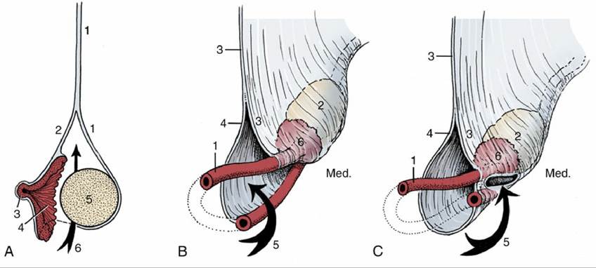
FIG. 5.60 (A) Schematic representations of the ovary and the suspensory system of the ovary and uterine tube and of the varying forms of the ovarian bursa. 1, Mesovarium; 2, mesosalpinx; 3, abdominal opening of uterine tube; 4, infundibulum; 5, ovary; 6, arrow is in the ovarian bursa. (B) Spacious bursa with large entrance (cow, mare). (C) Bursa with constricted entrance and entrapped ovary (bitch). 1, Uterine tube; 2, ovary; 3, mesovarium; 4, mesosalpinx; 5, arrow entering the ovarian bursa; 6, infundibulum; Med., medial.
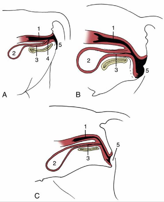
FIG. 5.61 Variation in the position of the vestibule in relation to the ischial arch in the (A) cow, (B) mare, and (C) bitch.
1, Vagina; 2, bladder; 3, urethra; 4, suburethral diverticulum; 5, vulva.
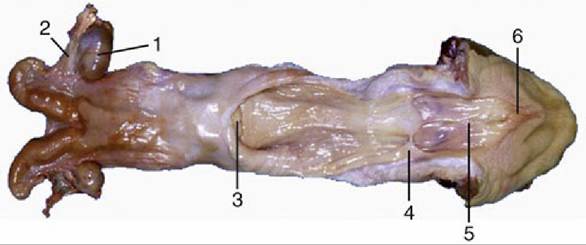
FIG. 5.62 Uterus and opened vagina of the cow. 1, Ovary; 2, uterine tube; 3, cervix; 4, hymen; 5, vestibule; 6, glans of clitoris.
When followed distally from its attachment to the abdominal roof, the mesovarium, which supports the ovary, releases a lateral fold (mesosalpinx) that passes onto the uterine tube (Figs. 5.58/7 and 5.60A). Mesosalpinx and mesovarium enclose a pouch, the ovarian bursa, into which the ovary projects. The bursa may be shallow and unable to hold the ovary (mare; Fig. 5.58/9) or deep and so enclosed by the fusion of apposed serosal surfaces that the ovary is permanently trapped (bitch; Fig. 5.60C). In certain nondomestic species (e.g., mouse) fusion is so complete that the space within the bursa no longer communicates with the peritoneal cavity. The walls of the bursa may contain so much fat that the ovary is quite hidden. The mesovarium also supports a fibromuscular band, the proper ligament of the ovary, which extends from the caudal pole of the ovary to the adjacent tip of the horn of the uterus.
The large part of the broad ligament that passes onto the horn and body of the uterus helps to give the organ the shape characteristic of the species. The two serosal membranes are very widely separated by fat where they attach to the cervix and especially to the vagina and make the lateral part of the vagina retroperitoneal. A cord of fibrous tissue and smooth muscle, the round ligament of the uterus, passes from the tip of the horn of the uterus toward (and in the bitch, through) the inguinal canal, supported by a special fold of peritoneum detached from the lateral surface of the broad ligament.
The muscles and fasciae associated with the female reproductive organs are best considered in topographical contexts for those animals such as ruminants in which they have special importance (p.
692). It will be recalled that the pelvic outlet is closed by a musculofascial partition of complicated form and structure. The dorsal part, the pelvic diaphragm, closes the outlet about the anus. The ventral part, the urogenital diaphragm (membrana perinei), closes the outlet about the vestibule. Muscle forms the principal component of the pelvic diaphragm, whereas the fasciae predominate in the urogenital diaphragm.The blood supply to the female reproductive organs is obtained from several sources. The ovarian artery, a direct branch of the aorta, supplies the ovary and branches in varying patterns to the uterine tube and cranial part of the horn of the uterus. The ovarian artery assumes an extraordinarily convoluted course and, depending on species, is more or less closely related to the ovarian vein. The uterine branch anastomoses with the uterine artery within the broad ligament (Fig. 5.63/1' and 2).
The uterine artery arises as an indirect branch of the internal iliac artery (except in the mare) and runs forward within the broad ligament. It detaches a series of anastomosing branches to the body and horn of the uterus; the most cranial anastomoses with the ovarian artery, the most caudal with the vaginal artery. Thus, an arterial arcade is established, running the length of the uterus and supplied from both ends (Fig. 5.64). It is thought that more blood supply may occur to areas such as implantation sites in the uterus, and a reduced supply to certain parts may result in runts, which are so common in pigs. The more caudal parts of the tract are variously supplied by branches of the internal pudendal and vaginal arteries; some more important species differences are mentioned elsewhere.
The veins, broadly satellite to the arteries. The plexiform ovarian vein is relatively much larger, the uterine vein relatively much smaller, than the accompanying artery (Fig. 5.63). A prominent and elaborate venous plexus present on the ventral aspect of the uterus and vagina drains both organs via any of the paired ovarian, uterine, and vaginal veins.
The close relation between the artery and ovarian vein, best seen in ruminants and sows, provides a means for the countercurrent transfer of the luteolytic hormone (prostaglandin) from venous to arterial blood (p. 198).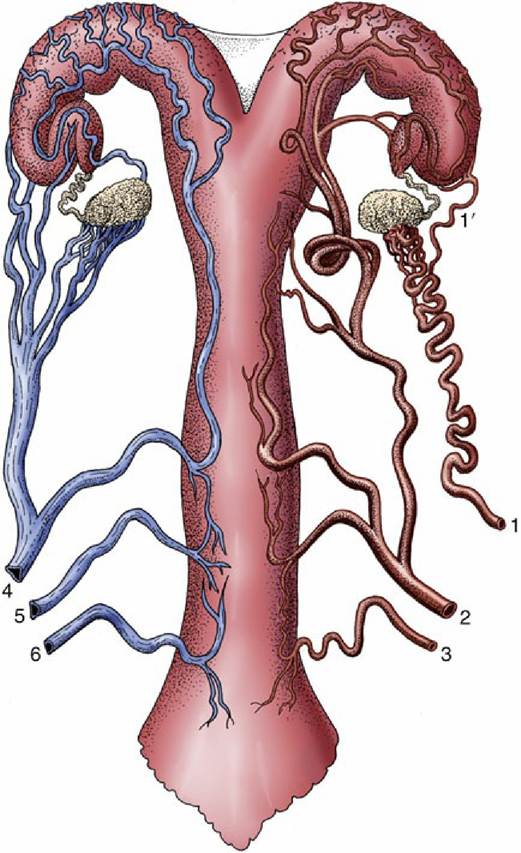
FIG. 5.63 Semischematic ventral view of the blood supply to the reproductive tract of the cow. The arteries are depicted on the right side, the veins on the left. 1, Ovarian artery; 1', uterine branch; 2, uterine artery; 3, vaginal artery; 4, ovarian vein; 5, accessory vaginal vein; 6, vaginal vein.
The lymphatics from the ovaries and more cranial parts of the tract pass to the aortic and medial iliac nodes, and those from more caudal parts to the medial iliac and other nodes within the pelvis.
Innervation of the female reproductive organs is provided by both sympathetic and parasympathetic fibers, by routes that have yet to be fully clarified. Although sympathetic fibers run to the ovary including the ripening follicles together with the ovarian artery, their denervation hardly disturbs ovarian function. The fibers to the uterine tube, uterus, and vagina mainly follow the other arteries to form plexuses within the broad ligaments and the genital organs themselves. In the caudal part of the broad ligaments these fibers are augmented by other sympathetic fibers that travel by way of the plexus located in the retroperitoneal pelvic tissue.

FIG. 5.64 Semischematic drawing of the blood supply of the female reproductive tract (bitch). 1, Ovarian artery; 2, uterine branch of the ovarian artery; 3, vaginal artery; 4, uterine artery.
The parasympathetic fibers branch from the pelvic nerves and reach the genital organs via the pelvic plexus. A large proportion goes to erectile tissue.
Both sympathetic and parasympathetic fibers seem to be concerned with uterine activity, although their precise roles in stimulation and inhibition are still controversial. The uterus is able to coordinate contractions and accomplish a normal birth even after denervation.