The anatomy of the female reproductive organs is strongly influenced by age, present status, and previous reproductive history.
The initial description refers to the mature, parous but nongravid mare (Fig. 22.9).
1
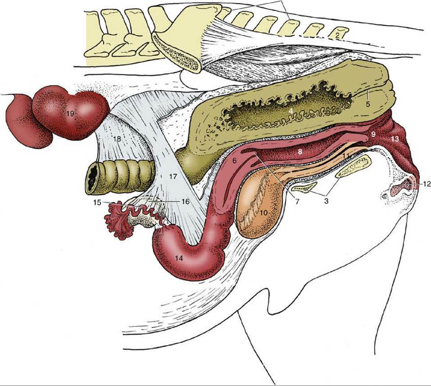
FIG. 22.8 Caudal abdominal and pelvic organs of the mare in situ; the organs have been sectioned in a paramedian plane with the pelvis. Because of the absence of the intestines, the ovaries hang much lower than they would in the intact animal. 1, Sacrum; 2, caudal vertebra 2; 3, floor of pelvis; 4, rectum; 5, anal canal; 6, cervix; 7, vaginal part of cervix; 8, vagina; 9, vestibule; 10, bladder; 11, urethra; 12, clitoris; 13, vulva; 14, left uterine horn; 15, uterine tube; 16, ovary; 17, broad ligament (largely cut away); 18, descending mesocolon; 19, left kidney.
More on the topic The anatomy of the female reproductive organs is strongly influenced by age, present status, and previous reproductive history.:
-
Veterinarian -