» The Bladder and Female Urethra
The neck region of the bladder lies directly on the pelvic floor, and when the organ is fully contracted it forms a firm, globular swelling about the size of a clenched fist in the pelvic cavity and becomes wholly retroperitoneal.
As the bladder fills, it gradually assumes a more ovoid form and extends cranially into the abdomen.The relations of the bladder depend on the degree of filling and on the sex. When empty, its vertex is generally in contact with the pelvic flexure of the colon, but as the bladder enlarges, the vertex and adjacent parts obtain a more extensive and more varied relationship to the intestine. In the mare the dorsal surface is in contact with the cranial part of the vagina, the cervix, a variable part of the body of the uterus, and sometimes the rectum (Fig. 22.8). The corresponding relations in the male are the genital fold, the deferent ducts, the vesicular glands, the prostate, and the rectum.
The relatively large neonatal bladder is entirely intra-abdominal and adjusts to adult proportions and position with the postnatal development of the pelvis and the intestines. Leakage at the navel from a still-patent urachus is not uncommon in the first period after birth and provides a potential portal for infection.
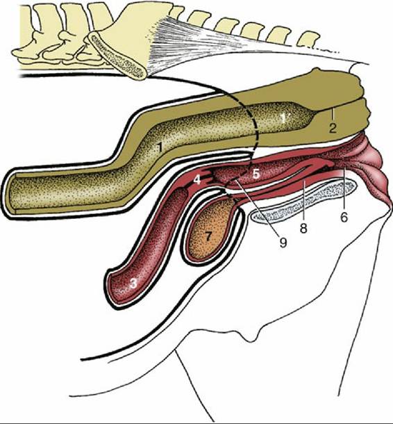
FIG. 22.5 Median section of the pelvis of the mare (schematic). 1 and 1', Peritoneal and retroperitoneal parts of the rectum, respectively; 2, anal canal; 3, uterus; 4, cervix; 5, vagina; 6, vestibule; 7, bladder; 8, urethra; 9, caudal extent of peritoneum.
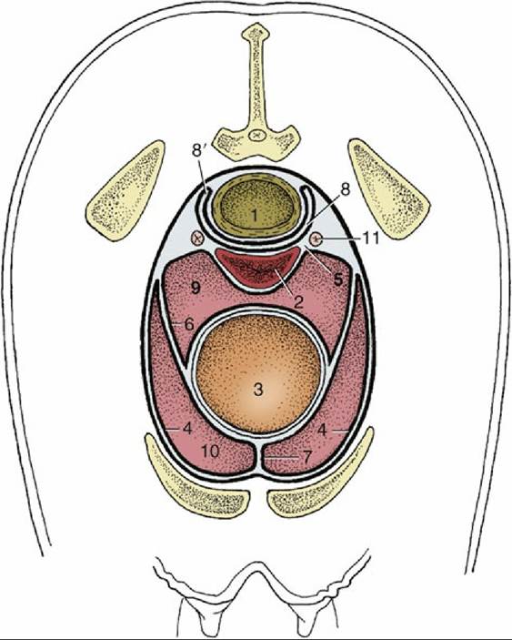
FIG. 22.6 The disposition of the peritoneum in the pelvis of the mare (transverse section). 1, Rectum; 2, vagina; 3, bladder; 4, parietal peritoneum; 5, broad ligament; 6, lateral ligament of bladder; 7, median ligament of bladder; 8, rectogenital pouch; 8', pararectal fossa; 9, vesicogenital pouch; 10, pubovesical pouch; 11, ureter.
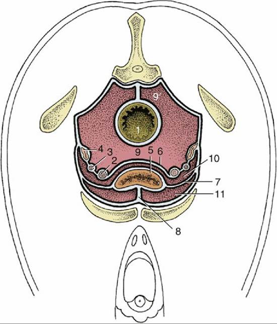
FIG.
22.7 The disposition of the peritoneum in the pelvis of the stallion (transverse section). 1, Rectum;2, deferent duct; 3, ureter; 4, vesicular gland; 5, bladder; 6, genital fold; 7, lateral ligament of bladder; 8, median ligament of bladder; 9, rectogenital pouch; 9', pararectal fossa; 10, vesicogenital pouch; 11, pubovesical pouch.
Surgical access to the urinary bladder is needed for removal of urinary calculi (cystotomy), to repair a disrupted bladder (cystorraphy), and to repair a patent or persistent urachus (cystoplasty). In the mare, an incision of 6 to 8 inches is made beginning slightly cranial to the umbilicus and extending caudally. In the male, the incision goes paramedially around the prepuce.
The female urethra is very short (only 6 cm or thereabouts) and opens into the vestibule, immediately caudal to the transverse fold of the hymen. It wide enough to admit one finger without difficulty, and a small hand by gentle manipulation and low epidural anesthesia, which is convenient when returning a bladder prolapse or removing a urolith from the bladder. The shortness, wide caliber, and dilatable nature of the urethra permit occasional prolapse of the bladder into the vestibule.
The male urethra is described with the reproductive organs.