The applied importance of the lymphatic drainage has been stressed, and accounts of its organization in different species are presented later.
Because these accounts are necessarily fragmented by the regional character of the later chapters, it may be useful to give a short general account here. We begin with Figs. 7.53 and 7.54, which show the palpable lymph nodes of the dog and the cat.
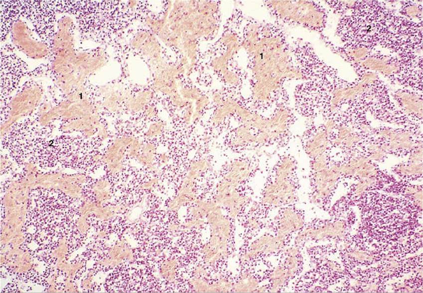
FIG. 7.49 Hemal node of sheep (hematoxylin and eosin; magnification ?70). 1, Erythrocytes; 2, lymphocytes.
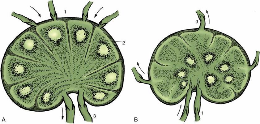
FIG. 7.50 (A) Structure of a lymph node in which the germinal centers (lymph nodules) occupy the cortical region. (B) In the pig the germinal centers lie centrally. Arrows indicate the direction of lymph flow. 1, Afferent lymphatics; 2, subcapsular sinus; 3, efferent lymphatics.

FIG. 7.51 Lymph node (dog) (magnification ?28). 1, Cortex with lymph nodules; 2, medulla; 3, afferent
lymph vessels.
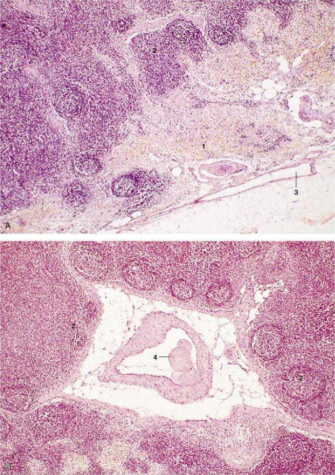
FIG. 7.52 (A) and (B) Lymph node (pig) (magnification ?28). 1, Loose Iymphoreticular tissue; 2, lymph nodules in centrally located “cortex”; 3, efferent lymph vessels; 4, centrally located afferent lymph vessel, with valve.
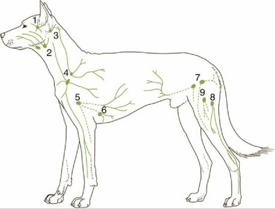
FIG. 7.53 Palpable lymph nodes of the dog. 1, Parotid; 2, mandibular; 3, lateral retropharyngeal (inconstant); 4, superficial cervical; 5, axillary; 6, accessory axillary (inconstant); 7, superficial inguinal; 8, popliteal; 9, femoral (inconstant).
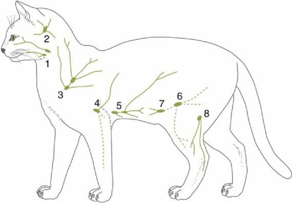
FIG. 7.54 Palpable lymph nodes of the cat.
1, Mandibular; 2, lateral retropharyngeal; 3, dorsal superficial cervical; 4, axillary; 5, accessory axillary; 6, superficial inguinal; 7, caudal epigastric; 8, popliteal.The Lymph Nodes of the Head
Three Iymphocenters are present in the head. The parotid center consists of one or more nodes placed on the masseter close to the temporomandibular joint and commonly covered by the parotid gland (Fig. 7.55/2). These nodes receive lymph from dorsal structures of the head, including skin, the dorsal bones of the skull, the contents of the orbit, and the masticatory muscles (in part).
The mandibular center (Fig. 7.55/1) comprises a group of nodes placed within the intermandibular space or more caudally by the angle of the jaw. They drain structures of the muzzle, the salivary glands, the intermandibular space (including the tongue), and a further part of the masticatory muscles.
The retropharyngeal center consists of two groups of nodes, medial and lateral; the former (Fig. 7.55/4) lie against the roof of the pharynx, and the latter (Fig. 7.55/3) are contained within the atlantal fossa. Together, they drain deeper structures of the head and adjacent parts of the neck, including the pharynx and larynx; one or the other also receives lymph that has already passed through the more peripheral centers. In most species the medial group receives the output from the lateral retropharyngeal, parotid, and mandibular nodes; in cattle this role is taken by the lateral group (see in Chapter 25).
The Lymph Nodes of the Neck
The superficial cervical center (Fig. 7.55/6) lies in front of the shoulder, under cover of the lateral superficial muscles of the neck. It consists of one or more nodes that drain a very wide but predominantly superficial territory. It extends from the nape to the middle of the trunk and includes the proximal part of the forelimb. The outflow is usually to the lymphatics at the thoracic inlet (Fig. 7.55/12).
The deep cervical center (Fig. 7.55/5) comprises a chain of nodes usually arranged in cranial, middle, and caudal groups but often irregular in disposition.
The nodes are placed along the trachea within the visceral space of the neck and mainly drain deeper and more ventral structures. Much of this lymph percolates through successive nodes of the chain before entering one of the major lymphatic channels at the entrance to the chest.The Tracheal Duct
In most species the tracheal duct (Fig. 7.55/12) is a large paired vessel that follows the course of the trachea within the neck. Except in the horse, it takes origin in the retropharyngeal nodes that serve as the collecting center of the head. It may be augmented by tributaries from deep cervical nodes before it joins the thoracic (on the left side) or right lymphatic duct. Alternatively, one or both tracheal ducts may enter the corresponding jugular or other vein at the venous confluence at the entrance to the thorax (see Fig. 1.34). In the horse the flow may be interrupted by serial passage through deep cervical nodes (see in Chapter 18).
The Lymph Nodes of the Forelimb
One axillary center exists. The principal nodes are contained within the axilla where they lie on the medial muscles of the shoulder. Additional nodes may be found in relation to the first rib or more caudally on the chest wall. In the horse alone, a more distal group of cubital nodes is placed over the medial aspect of the elbow. The center drains the deeper structures of the entire limb and the more superficial structures of the distal segments, and the collection goes to one of the major lymphatic or venous channels at the entrance to the chest.
The Lymph Nodes of the Thorax
Four lymphocenters attend to the drainage of the thoracic walls and contents. The nodes within certain groups are rather diffusely spread, and it is not always easy to decide their correct designation.
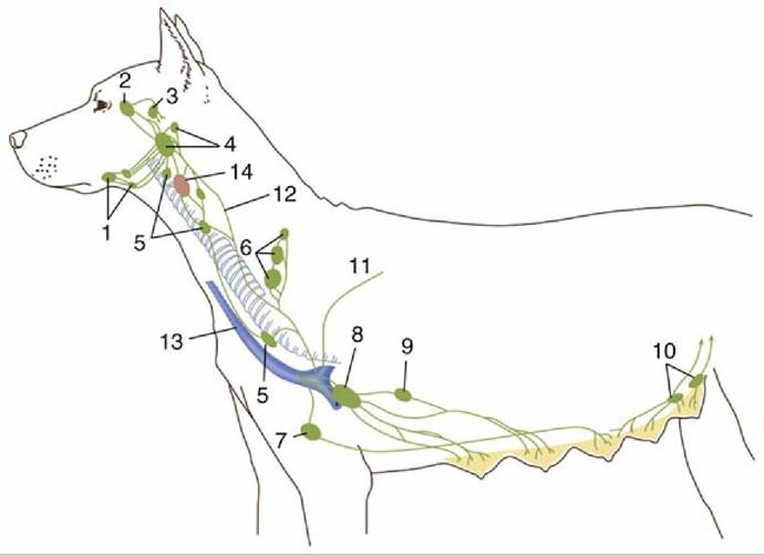
FIG. 7.55 Lymph drainage of the head, neck, and mammary glands of the dog. 1, Mandibular nodes; 2, parotid node; 3, lateral retropharyngeal node; 4, medial retropharyngeal nodes; 5, cranial and caudal deep cervical nodes; 6, superficial cervical nodes; 7, sternal node; 8, axillary node; 9, accessory axillary node;
10, superficial inguinal nodes; 11, thoracic duct; 12, tracheal duct; 13, external jugular vein; 14, thyroid gland.
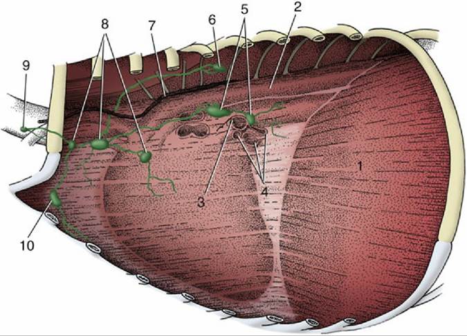
FIG. 7.56 Thoracic lymph nodes in the dog. Left lung removed; the outline of the heart is visible within the mediastinum. 1, Diaphragm; 2, thoracic aorta; 3, left bronchus; 4, pulmonary vessels; 5, tracheobronchial nodes; 6, intercostal node; 7, thoracic duct; 8, cranial mediastinal nodes; 9, caudal deep cervical node; 10, sternal node.
The dorsal thoracic center comprises two groups of small, inconstant nodes. The intercostal set (Fig. 7.56/6) is found within the upper parts of a few intercostal spaces. The thoracic aortic set is dispersed along the course of the vessel. The center drains the back and the deeper tissues of the thoracic wall and sends its outflow, possibly after serial passage through several nodes, to the thoracic duct or the mediastinal nodes (Fig. 7.56/8).
The ventral thoracic center consists of cranial sternal nodes (Fig. 7.56/10) by the manubrium of the sternum and, only in ruminants, caudal sternal nodes placed against both surfaces of the transversus thoracic muscle. The center drains the deeper structures of the ventral part of the thoracic wall and sends its efferent flow either to mediastinal nodes or to one of the larger collecting vessels.
The mediastinal center is divided into a group of nodes within the cranial mediastinum (Fig. 7.56/8), a middle group about the base of the heart, and a caudal group (absent in carnivores) near the esophagus as it approaches the diaphragm (see in Chapter 27). The various nodes drain structures of the thoracic wall, mainly after first passage of the lymph through other primary nodes, and thoracic viscera. They provide a secondary station for lymph from the lungs that has already passed through tracheobronchial nodes. The outflow goes to the large collecting vessels at the entrance to the chest, in part after serial passage through several nodes.
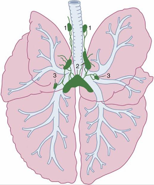
FIG.
7.57 Lymph nodes associated with trachea and lungs of the dog. 1, Cranial mediastinal nodes; 2, tracheobronchial nodes; 3, pulmonary nodes.The bronchial center consists of groups of tracheobronchial nodes placed about the tracheal bifurcation and, in many animals, small pulmonary nodes embedded within the substance of the lung (Figs. 7.56/5 and 7.57). The former groups are individually named (left, middle, right, and [in ruminants and pigs] cranial tracheobronchial nodes) according to their relationships to the major bronchi. They collect lymph from the lungs and send it in inconstant fashion to middle and caudal mediastinal nodes and sometimes directly to the thoracic duct.
The Thoracic Duct
The thoracic duct is the major lymph-collecting channel. It arises from the cisterna chyli, which receives lymph from the abdomen, pelvis, and hindlimbs (see Fig. 1.34/5 and 7). The cisterna has a very irregular, even plexiform, shape, and although it is mainly contained between the aorta and the vertebrae at the thoracolumbar junction, it may also extend ventrally around the vena cava and the origin of the celiac artery. The thoracic duct passes through the aortic hiatus into the mediastinum. Its further course takes it cranially and ventrally, over the left face of the trachea, to a termination within one of the veins, most often the left jugular vein, that form the cranial vena cava or the vena cava itself (Fig. 7.58). The duct receives additional lymph from the structures and nodes of the left side of the chest. A separate right lymphatic duct provides similar drainage for cranial thoracic structures of the right side and proceeds to a similar termination. One or both commonly receive the corresponding tracheal duct(s).
FIG. 7.58 Lymphangiogram of the canine thoracic duct.
The Lymph Nodes of the Abdominal Viscera and Loins
The roof of the abdomen is drained by a lumbar center containing various nodes spread along the abdominal aorta and possibly also within the spaces between the lumbar transverse processes (Fig.
7.59). Usually those (renal) nodes (Fig. 7.59/7) that are associated with the kidneys are larger than others in the series. In addition to draining the structures of the loins, kidneys, and adrenal glands, these nodes may receive some lymph from reproductive organs. The flow is to the cisterna chyli (Fig. 7.59/5) directly or after serial passage.Three centers associated with the drainage of the abdominal viscera have territories broadly corresponding to those of the celiac, cranial mesenteric, and caudal mesenteric arteries. They show very considerable interspecific distinctions and include the following (Fig. 7.60). The celiac center comprises splenic, gastric (subdivided in ruminants), hepatic, and pancreaticoduodenal nodes (Fig. 7.60/1-4). The cranial mesenteric center consists of cranial mesenteric nodes toward the root of the mesentery and more peripheral jejunal, cecal, and colic nodes (Fig. 7.60/5-7). The caudal mesenteric center comprises caudal mesenteric nodes associated with the descending colon (Fig. 7.60/8). The three centers give rise to various visceral trunks that converge on the cisterna chyli.
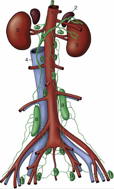
FIG. 7.59 Lymph drainage of the canine lumbosacral area, ventral view. 1, Aorta; 2, adrenals; 3, kidneys; 4, caudal vena cava; 5, cisterna chyli; 6, lumbar aortic nodes; 7, renal nodes; 8, medial iliac nodes; 9, hypogastric nodes; 10, sacral nodes; 11, deep inguinal (iliofemoral) nodes.
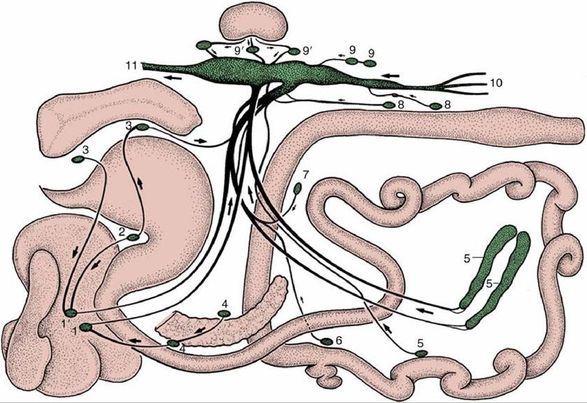
FIG. 7.60 Lymph drainage from the organs in the canine abdominal and pelvic cavities (schematized). 1 and 1', Right and left hepatic nodes; 2, gastric node; 3, splenic nodes; 4, pancreaticoduodenal nodes; 5, jejunal nodes; 6, right colic node; 7, middle colic node; 8, caudal mesenteric nodes; 9, lumbar aortic nodes; 9', renal nodes; 10, efferents from the iliosacral region; 11, continuation of cisterna chyli as thoracic duct.
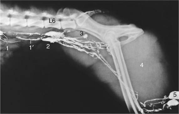
FIG. 7.61 Lymphangiogram of the canine lumbar area, pelvis, and thigh. 1, Lumbar aortic lymph node; 1', lumbar trunks; 2, medial iliac nodes; 3, hypogastric node; 4, thigh muscles; 5, popliteal nodes; L6, sixth lumbar vertebra.
The Lymph Nodes of the Hindlimb, Pelvis, and Abdominal Wall
Although an inconveniently large territory to consider together, the hindlimb, pelvis, and abdominal wall cannot be subdivided because the responsibilities of certain nodes do not coincide with the usual division of the body. The most peripheral popliteal center, which consists of a node (or nodes) placed within the popliteal fossa caudal to the stifle (Figs. 7.53/8 and 7.61/5), drains the distal part of the limb. The efferent flow is directed to the medial iliac center (except in the horse, in which it passes to deep inguinal nodes).
The ischial center contains the ischial node placed on the lateral aspect of the sacrosciatic ligament (of ungulates [see in Chapter 31]-no comparable node exists in carnivores). It collects from the muscles and skin of the rump and proximal thigh and sends its outflow to various nodes of the iliosacral center.
The nodes of the deep inguinal (iliofemoral) center are placed along the course of the external iliac artery or its femoral continuation (Fig. 7.59/11). They primarily drain part of the thigh but also accept lymph from the popliteal nodes for onward passage to the iliosacral center.
The more peripheral superficial inguinal center includes the superficial inguinal nodes of the groin, the subiliac nodes of the flank fold (except in the dog), the coxal node, and the nodes of the paralumbar fossa of cattle (Figs. 7.55/10 and Chapter 31). The superficial inguinal nodes are also named scrotal or mammary because they drain the external male reproductive organs or the udder (in dogs, caudal mammary glands) in addition to the groin region. The subiliac node drains skin and deeper structures extending from the midflank to the thigh. The efferent lymph passes to the iliosacral center, directly or after passage through the deep inguinal nodes.
The iliosacral center is a very large, widely spread collection of nodes placed against the roof of the caudal part of the abdomen and within the pelvic cavity (see Fig. 7.59). The main components are the medial iliac nodes (Fig. 7.59/8), near the origin of the external and internal iliac arteries and, though not in the dog, the lateral iliac about the branching of the deep circumflex iliac vessels. There are other nodes on the walls (sacral nodes) and about the viscera (hypogastric and anorectal nodes) in the pelvic cavity. These various small nodes are the primary filtration centers for adjacent structures and secondary stages in the drainage of the hindlimb and reproductive and other pelvic organs. The lymph from these nodes flows to the medial iliac nodes that give origin to the lumbar trunks.
The Lumbar Trunks
The lumbar trunks are formed mainly by efferent vessels from the medial iliac nodes. They form a plexus on the roof of the abdomen, where they are augmented by part of the lumbar outflow before they expand as the cisterna chyli (Figs. 7.59/5 and 7.61/1'). This structure also receives visceral trunks from the digestive organs.