The spleen* is contained within the left cranial part of the abdomen, where it is joined to the greater curvature of the stomach by inclusion within the greater omentum.
The spleen's precise position depends on the degree of filling of the stomach and on its own blood content. The basic form is very dissimilar in the various domestic species, being dumbbell-shaped in the dog and cat, straplike in the pig, a broader oblong shape in cattle, and falciform in the horse (Fig.
7.62). Its capsule extends trabeculae into the interior. In some species (carnivores) the capsule and trabeculae are very muscular, in others (ruminants) much less so, and these differences determine the extent of the physiologic variation in size. When relaxed, the spleen of the dog and cat increases many times from its contracted state.The soft tissue contained within the supporting framework is divided between red and white pulp. Red pulp consists of spaces in series with the blood vessels and is occupied by a concentrated cellular elements of the blood. The white pulp, which is divided into foci that are usually just visible to the naked eye, is formed of lymph nodules within a supporting reticuloendothelial framework. This tissue has the usual lymphogenic and phagocytic properties.
The functions of the spleen are blood storage, the removal of particulate matter from the circulation, the destruction of worn-out erythrocytes, and the production of lymphocytes. The blood storage function is familiar to all who have experienced a "stitch in the side,'' the pain that sometimes accompanies physical stress and is associated with contraction of the splenic capsule.
The spleen is supplied by the generously sized splenic artery, a branch of the celiac artery (see Fig. 3.39/4). The venous drainage through the splenic vein leads to the portal vein (see Fig. 3.50/1 and 2). Important specific features in the arrangement of these vessels exist. The artery and vein may pass undivided through a confined hilus (ruminants; Fig. 7.62B); may run the length of the organ, detaching branches at intervals (horse, pig; Fig. 7.62A); or may divide as they approach the spleen into branches that vascularize splenic compartments that are normally independent, although they do communicate (dog, cat; Fig. 7.62C). The lymph vessels found in the capsule and trabeculae do not extend into the pulp. The sympathetic and parasympathetic nerves approach with the artery.
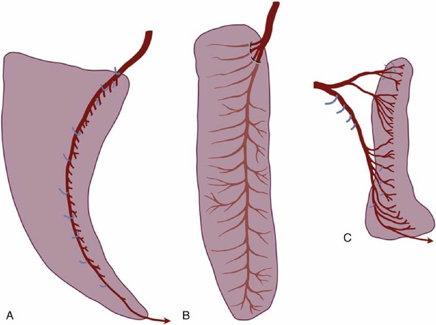
FIG. 7.62 Visceral surfaces of the spleens of (A) horse, (B) cattle, and (C) dog to show the distribution of the splenic arteries. Branches to other structures are shown in blue.
The spleen develops from a mesodermal condensation within the dorsal mesogastrium (which becomes the greater omentum) (see Fig. 3.65/6). The part of the sheet intervening between the stomach and the spleen may be specifically distinguished as the gastrosplenic ligament.