The appropriate regulation of visceral activities involves both afferent and efferent functions.
Visceral afferent pathways, however, are in general indistinguishable in structure and arrangement from their somatic afferent counterparts. In contrast, the visceral efferent pathways are clearly distinguished from their somatic efferent pathways.
These distinctions include the existence of two neurons in series (the preganglionic, myelinated fiber and a postganglionic thinly myelinated fiber), the location of the last neuron in the chain within a peripheral ganglion, and the restriction of the location of the preganglionic cell bodies to specific nuclei of the brainstem and particular regions of the cord (Figs. 8.72 8.73, and 8.76). Thus the term autonomic nervous system was originally, and is still best, defined as wholly efferent. Moreover, certain anatomic, physiologic, and pharmacologic features distinguish the two contrasting divisions of the autonomic system-sympathetic and parasympathetic—whereas no similar distinction exists for visceral afferent pathways. Visceral afferent fibers are, nonetheless presumed to be included in all cranial and spinal nerves, if only because of the ubiquitous distribution of blood vessels.Before we move to more detailed descriptions of the specifics of the parasympathetic and sympathetic systems in the next section, one general distinction relates to the nature of the actions of the two systems—namely, that the activities of the parasympathetic system tend to be more discrete than those of the sympathetic system. Acetylcholine is used at the synapse between the postganglionic parasympathetic neuron and the target organ, and because acetylcholine is liberated and destroyed locally, its effects tend to be very specific. The narrower localization of parasympathetic responses is further assisted by the location of parasympathetic ganglia close by or even within the target organ. In contrast, norepinephrine is used as the neurotransmitter at the last synapse of the sympathetic pathway except where epinephrine is produced by the adrenal medulla, from which it is released into the bloodstream, evoking a mass sympathetic response.
Additionally the sympathetic ganglia are located closer to the central nervous system, such that sympathetic postganglionic fibers radiate more widely before reaching their target organ, resulting in more general and less discrete sympathetic responses.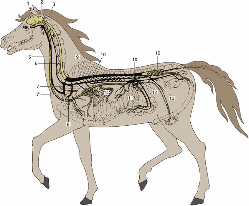
FIG. 8.76 Distribution of sympathetic (black) and parasympathetic (dotted yellow) nervous systems, semischematic. 1, Parasympathetic oculomotor nucleus; 2, salivatory nuclei (rostral and middle parasympathetic nuclei); 3, dorsal vagal nucleus; 4, cranial cervical ganglion; 5, vagosympathetic trunk; 6, vertebral nerve; 7, cervicothoracic ganglion; 7', middle cervical ganglion; 8, ansa subclavia; 9, sympathetic outflow from spinal cord; 10, sympathetic trunk with paravertebral ganglia; 11, celiac ganglion; 12, cranial mesenteric ganglion; 13, caudal mesenteric ganglion; 14, vagus nerve with distribution to thoracic and abdominal organs; 15, sacral outflow of parasympathetic nervous system.