The Arterial Blood Supply
The blood supply to the brain comes mainly from the circulus arteriosus cerebri (formerly known as the circle of Willis), which lies ventral to the hypothalamus, where it forms a ring around—but at some distance from—the infundibular stalk.
The appearance of the circle and the pattern of its major branches are remarkably constant among mammals, although the arterial sources that supply the circle and the directions in which blood flows in certain vessels vary among species. For this reason, the initial description given here is based on the arrangements in the dog, which are not only relatively simple but also the most common.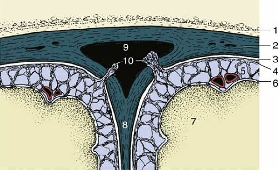
FIG. 8.64 Transverse section of the dorsal sagittal sinus and adjacent meninges. Cerebrospinal fluid is transferred from the subarachnoid space to the sinus via the arachnoid granulations (villi). 1, Roof of cranial cavity; 2, fused dura mater and periosteum; 3, subdural space; 4, arachnoid; 5, subarachnoid space; 6, pia mater; 7, cerebral hemisphere; 8, falx cerebri; 9, dorsal sagittal sinus; 10, arachnoid granulations (villi).
The arterial circle of the dog is supplied from three sources: paired internal carotid arteries laterally and the basilar artery caudally (Fig. 8.65). The internal carotid artery (Fig. 8.65/5) arises as a terminal branch of the common carotid at the level of the pharynx. The internal carotid then travels toward the base of the skull. In many species the artery makes immediate entry to the cranial cavity through a carotid foramen in the cranial floor, but in the dog it must first traverse a tunnel (carotid canal) in the bone medial to the tympanic bulla. The artery leaves the rostral end of the tunnel and forms a loop that first carries it ventrally, then dorsally, before it finally enters the cranial cavity.
It then penetrates the outer meninges, a process that involves passage through the cavernous venous sinus enclosed within the dura, before dividing into rostral and caudal branches. The rostral branch of right and left internal carotid arteries unite across the midline to complete the rostral half of the circle. From this rostral portion, both the rostral and middle cerebral arteries arise. The caudal branch of each internal carotid artery anastomoses with the caudal communicating artery, a branch of the basilar artery (Fig. 8.65/6). The basilar artery originates farther caudally as single artery (Fig. 8.65/11 and see later) but divides into right and left branches at the level of the midbrain before each branch joins the internal carotid arteries to complete the circle. The caudal cerebral and rostral cerebellar arteries leave the caudal half of the circle; the fifth major artery to the brain, the caudal cerebellar, leaves the single basilar artery directly (Fig. 8.65/10).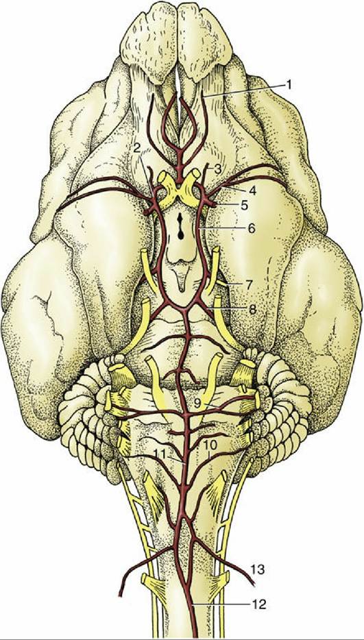
FIG. 8.65 Arteries on the ventral surface of the canine brain. 1, Internal ethmoidal artery (a.); 2, rostral cerebral a.; 3, internal ophthalmic a., 4, middle cerebral a.; 5, internal carotid a.; 6, caudal communicating a.; 7, caudal cerebral a.; 8, rostral cerebellar a.; 9, labyrinthine a.; 10, caudal cerebellar a; 11, basilar a.;
12, ventral spinal a.; 13, vertebral a.
The blood within the basilar artery has a composite origin. The artery appears to be the direct continuation of the small ventral spinal artery but is greatly reinforced by anastomosis with the vertebral artery (Fig. 8.65/13), which passes into the vertebral canal through the atlas. The vertebral artery itself receives anastomotic branches (dog and horse) from the occipital artery (another branch of the common carotid) before entering the canal, and it would thus appear that the occipital artery also contributes to the supply of the brain.
However, the vertebral artery is the main if not sole supply to the occipital lobes of the cerebral hemispheres and other caudal parts of the brain.The arrangement is more complicated in many other species (Fig. 8.66). In these, the internal carotid connects with other arteries of the head, especially the maxillary, before joining the arterial circle. The internal carotid at this level may be a small vessel initially, but in many species it enlarges closer to the brain and detaches many tortuous branches, which then rejoin to form the original single channel before meeting up with the arterial circle. This arrangement, which may present a rather tangled appearance, is known as a rete mirabile and has a rather enigmatic significance; the arrangement enhances the efficiency of the blood-cooling mechanism that is discussed shortly. In some species, the lumen of the part of the internal carotid artery proximal to the rete becomes obliterated, sometimes only a considerable time after birth; when this happens, the distal artery from the rete that delivers blood to the brain is wholly of external carotid origin (see Fig. 7.36). This arrangement is found in both sheep and cattle, although these species differ in other features of the arterial supply to the brain (p. 648).
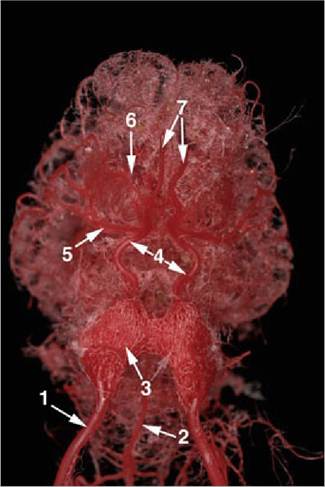
FIG. 8.66 Corrosion cast of the pig's brain (ventral view). 1, Internal carotid artery; 2, Basilar artery; 3, Rete mirabile; 4, cerebral arterial circle; 5, middle cerebral artery; 6, rostral cerebral artery; 7, internal ethmoidal artery.
The brain, particularly its gray substance, has very high metabolic requirements, and the arterial supply is commensurate with them, amounting to 15% or 20% of the cardiac output. Nevertheless, the vessels that actually penetrate the brain are uniformly small, a feature that may be related to the need to avoid large, pulsating arteries within the delicate brain tissue.
Moreover, in sharp contrast to the wide anastomoses between the large vessels supplying the brain, any intracerebral anastomoses are narrow and mostly connect functional end-arteries. This fact, coupled with the very limited regeneration capacity of brain tissue, explains why occlusion or rupture of a small sole vessel, being the only effective blood supply to some vital nucleus or tract, can have serious functional consequences. Notorious examples are provided by the small arteries within the human corpus striatum, where an infarct is often the cause of a stroke.The permeability of the blood capillaries of the nervous tissue is greatly reduced in comparison with that in other tissues, forming the blood-brain barrier. The main structural component of this barrier is composed of tight junctions between endothelial cells of brain capillaries, such that all substances to enter the brain tissue, other than small lipid-soluble molecules, must be transported through capillary endothelium. The blood-brain barrier is maintained through close cellular communication between brain endothelial cells and the pericytes and astrocytes surrounding these capillaries. There are several regions in the brain where the blood-brain barrier as described is not present; these regions, termed the circumventricular organs are described in detail elsewhere (Fig. 8.67) (p. 204).
The spinal cord is supplied by three arteries that run its craniocaudal length. The largest, the ventral spinal artery, follows the surface of the ventral fissure of the cord; paired dorsolateral spinal arteries run close to the furrow fissure from which the dorsal roots of the spinal nerves arise. All three vessels are periodically reinforced by branches from regional arteries: vertebral arteries in the neck and intercostal, lumbar, and sacral arteries in the trunk. These vessels enter at the intervertebral foramina, often in the form of narrow vessels that accompany the roots of the spinal nerves; they form plexuses on the surface of the cord with which the major longitudinal arteries connect.
This theoretically regular pattern is subject to much variation, both specific and individual, in which many expected reinforcing arteries are lacking, the plexus is unevenly developed, and stretches of the longitudinal trunks are attenuated.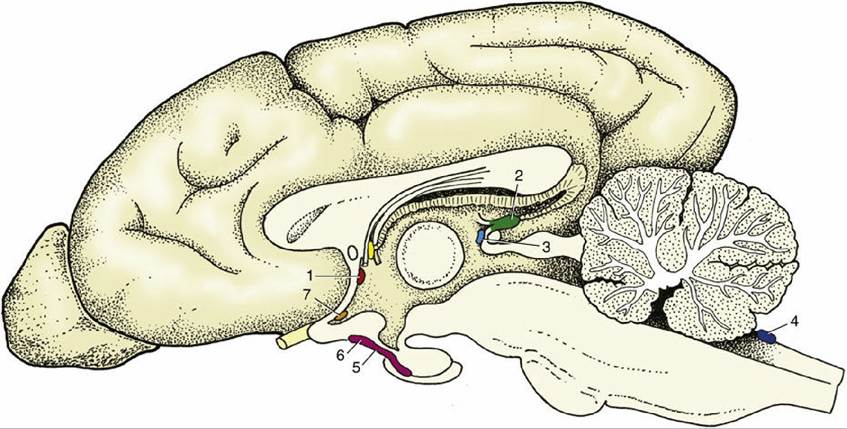
FIG. 8.67 Schematic median section of the canine brain with an indication of the locations of the Circumventricular organs. 1, Subfornical organ; 2, pineal body; 3, subcommissural organ; 4, area postrema; 5, posterior and intermediate lobes of pituitary; 6, median eminence; 7, vascular organ of lamina terminalis.
Branches of the ventral spinal artery supply the "core" of the cord, the gray substance, and the adjacent layer of white matter by an approach through the ventral fissure (see Fig. 19.5). The greater part of the white substance is supplied by radial branches from the dorsolateral arteries and surface plexus. Internal anastomoses between the two sets of vessels, although common, are of questionable efficiency.