The Meninges and Fluid Environment
The brain and spinal cord are surrounded by three continuous membranes or meninges composed of connective tissue that exhibit important topographic differences in their cranial and vertebral parts.
The tough outermost membrane, the dura mater, is fused with the inner periosteum of the skull bones; it splits from this within the margin of the foramen magnum to form a free tube of connective tissue separated from the wall of the vertebral canal by a distinct epidural space. The epidural space is occupied by fat, more fluid in life than in the postmortem specimen, and by the internal vertebral venous plexus; the fat and vessels together cushion the spinal cord and allow it to adjust to the movements of the neck and back (see Fig. 8.55). The dural tube is attached at its caudal end, where the several meninges finally combine in a fibrous strand (filum terminale) that fuses with the upper surface of the caudal vertebrae.
The fusion of the cranial dura with the periosteum obliterates the epidural space within the skull, and the cranial venous sinuses thus come to be enclosed within the thickness of the combined periosteum and dura. In addition to lining the cavity, the cranial dura forms certain folds that project inward and limit movements of the brain; these are a considerable hindrance to the removal of the intact brain at autopsy. One, the falx cerebri, extends ventrally from the dorsal and rostral cranial walls and serves to separate the two cerebral hemispheres; caudally the falx cerebri joins a second, transverse fold, the membranous tentorium cerebelli, which separates the cerebellum from the cerebrum (Fig. 8.57/7). The tentorium is ossified in its median part. A third specialization of the dura surrounds the dorsal aspect of the hypophysial fossa in which the hypophysis is seated, forming a diaphragm around the infundibular stalk.
A capillary space, the subdural space, divides the dura from the arachnoid, the first of the two more delicate inner meningeal membranes. This subdural space normally contains only a minute amount of a clear lymphlike fluid but may be enlarged by effusion of blood after an injury. The subdural space of the spinal cord is crossed by a bilateral series of triangular (denticulate) ligaments, each of which alternates with the origins of the spinal nerves; they attach the inner meninges to the dural tube and thus indirectly suspend the spinal cord within the dura along its craniocaudal length (Fig. 8.58/4). The outer part of the arachnoid forms a continuous membrane molded against the dural tube. The inner part of the arachnoid is composed of numerous fine trabeculae and filaments (imaginatively compared to a spider's web, hence the name arachnoid), which join the innermost meningeal membrane, the pia mater.
The pia mater covers the surface of the brain and spinal cord and follows every change in their contours. The pia mater is firmly attached to the outer surface of the brain and cord, and branches from arteries traveling within the pia penetrate the brain and spinal cord substance. These vessels are initially enclosed by pial sleeves, but as the vessels continue into the brain and spinal cord substance, the connective tissue of the pia soon merges with the vascular walls. In the spinal cord, a thickening of the pia fills the ventral fissure of the cord, where it appears as a glittering silver line.
All three meninges form cuffs around the roots of origin of the cranial and spinal nerves.
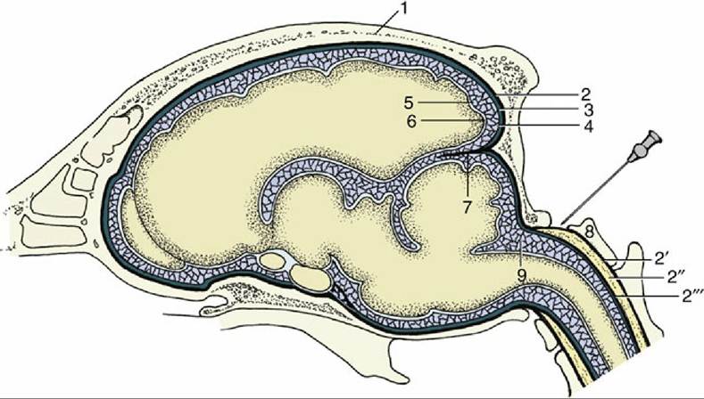
FIG. 8.57 Schematic representation of the meninges of the brain. The needle points to the atlanto- occipital space and the cerebellomedullary cistern. 1, Calvaria; 2, dura mater (also connected to the bone as periosteum); 2', periosteum of vertebral canal; 2", epidural space (with fat); 2'", dura mater of spinal cord; 3, subdural space; 4, arachnoid; 5, arachnoid space; 6, pia mater; 7, membranous tentorium cerebelli; 8, atlas; 9, cerebellomedullary cistern.
The subarachnoid space exists as the space between the pia and outer part of the arachnoid, traversed by the arachnoid trabeculae. The space containing the clear, watery cerebrospinal fluid is much wider than the subdural space but less uniform, particularly in its cranial part (see Fig. 8.57).
The widest parts ("cisterns") of the cranial subarachnoid space are located ventrally between the more prominent parts of the ventral surface of the brain and dorsally in the angle between the cerebellum and the dorsal aspect of the medulla. The dorsal widening, the cerebellomedullary cistern, is especially large and may be accessed in the living animal by a needle passed between the atlas and the skull through the foramen magnum (see Fig. 8.57). Cisternal puncture is employed in both clinical and experimental work for obtaining samples of cerebrospinal fluid. The spinal subarachnoid space is fairly uniform but widens around the conus medullaris, a fortunate circumstance because the subarachnoid space in the vertebral canal is accessed most easily by a dorsal route through the lumbosacral intervertebral space (Fig. 8.59).
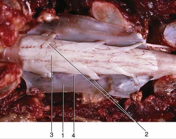
FIG. 8.58 Dorsal view of the opened vertebral canal. The dura mater has been dissected and is reflected. 1, Dura mater; 2, dorsal rootlets of a spinal nerve; 3, spinal cord (covered by pia mater); 4, denticulate ligament.
The cerebrospinal fluid within the subarachnoid space forms a water jacket that buoys up and protects the soft brain and cord. It is largely a product of the ependymal lining of the ventricular system within the brain, and most of the cerebrospinal fluid is produced at the choroid plexuses. The choroid plexuses are tufts of capillaries covered with ependyma that invaginate into the ventricles at specific locations throughout the brain (Fig. 8.60/6 and 9). An additional contribution to cerebrospinal fluid is made by the pial vessels.
The ventricles are the adult derivative of the lumen of the embryonic neural tube; the ventricles have complicated shapes, but because they are illustrated (Fig. 8.61) and the details have little veterinary significance, they need not be described. It is more important to understand their relationship to the choroid plexuses. The plexuses of each of the two lateral ventricles and of the third ventricle, which merge within the interventricular foramen, develop within a fold of pia that becomes entrapped between the expanding telencephalic vesicles and the roof of the diencephalon (Fig. 8.62). The plexuses of the fourth ventricle develop separately within the pia over the caudal medullary velum. In the course of development, these plexuses invaginate into the lumen of the fourth ventricle; parts later reemerge into the arachnoid space by herniating through paired lateral openings in the roof (Fig. 8.63).
The clear colorless cerebrospinal fluid is formed from the blood plasma by ultrafiltration through the "blood-cerebrospinal fluid barrier" at the choroid plexuses. Ependymal cells of the choroid plexuses are joined together with tight junctions, thus forcing any substances other than water or small lipophilic molecules to be transported through the cells to reach the ventricles. The fluid has a higher concentration of potassium and calcium ions and a lower concentration of sodium, magnesium, and chloride ions than the plasma; it is also rather deficient in glucose and, most important, contains little protein because the barrier is impermeable to larger molecules, which of course include those of many antibiotics and other drugs.
In addition to its mechanical role, the cerebrospinal fluid protects the brain through its chemical buffering capacity, which provides a rather stable milieu. It also transports nutrients, flushes away waste products, and serves as a medium for the diffusion of neuroendocrine and neurotransmitter substances.
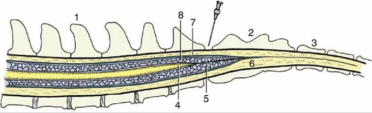
FIG.
8.59 Schematic median section of the vertebral canal and its contents. The needle points to the lumbosacral interarcuate space. 1, Lumbar vertebra; 2, sacrum; 3, caudal vertebra; 4, conus medullaris; 5, filum terminale; 6, epidural space; 7, dura mater; 8, arachnoid space with cerebrospinal fluid.
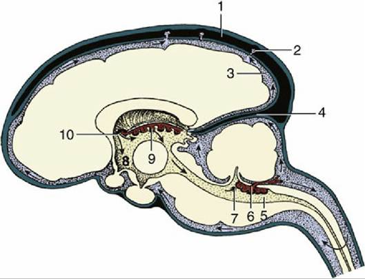
FIG. 8.60 The production and circulation of cerebrospinal fluid (sagittal section). The blood vessels are shown in black, the choroid plexuses are red, the subarachnoid spaces are stippled blue, the ventricles are stippled yellow, and the nervous tissue is solid yellow. The direction of the flow of the cerebrospinal fluid is indicated by arrows. The cerebrospinal fluid is secreted by the choroid plexuses (6, 9) of the lateral, third, and fourth ventricles. It escapes into the subarachnoid space via the aperture of the fourth ventricle (7). The cerebrospinal fluid is transferred to the systemic circulation (1) at the arachnoid villi (2). 1, Dorsal sagittal sinus; 2, subarachnoid space; 3, membranous tentorium cerebelli; 4, fourth ventricle; 5, choroid plexus of fourth ventricle; 6, aperture of fourth ventricle; 7, third ventricle; 8, choroid plexus of third ventricle; 9, interventricular foramen, connecting the lateral and third ventricles.
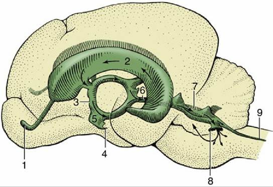
FIG. 8.61 Lateral view of a cast of the ventricles of the brain of the dog. 1, Cavity of olfactory bulb; 2, lateral ventricle; 3, third ventricle; 4, infundibular recess; 5, optic recess; 6, mesencephalic aqueduct; 7, fourth ventricle; 8, lateral recess; 9, central canal.
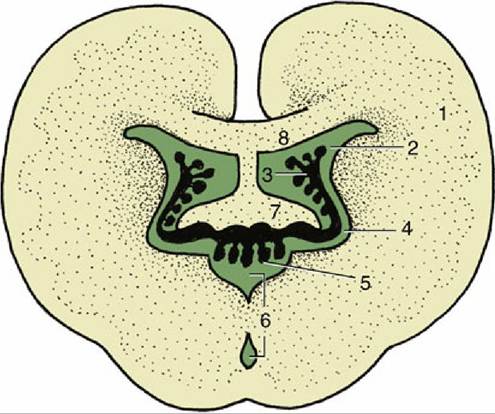
FIG. 8.62 Schematic section of the brain illustrating the interrelations of the third and lateral ventricles and their choroid plexuses. 1, Cerebral hemisphere; 2, lateral ventricle; 3, choroid plexus of lateral ventricle; 4,interventricular foramen; 5, choroid plexus of third ventricle; 6, third ventricle; 7, fornix; 8, corpus callosum.
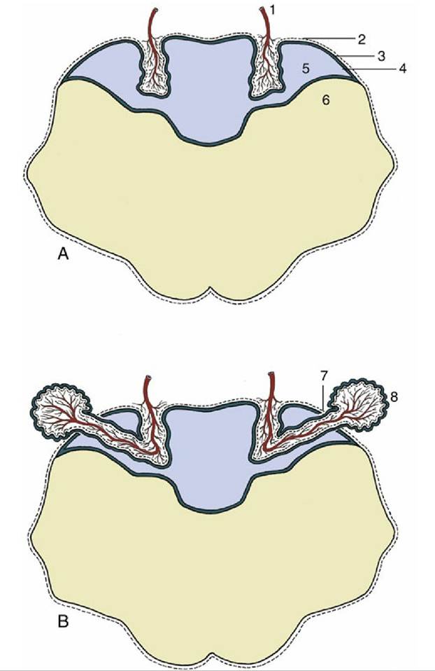
FIG. 8.63 (A) The formation of the choroid plexus in the roof of the fourth ventricle and (B) its later extension into the subarachnoid space. 1, Blood vessel invagination; 2, pia mater; 3, caudal medullary velum; 4, ependyma; 5, fourth ventricle; 6, myelencephalon; 7, aperture of fourth ventricle; 8, choroid plexus extending into subarachnoid space.
The fluid is produced continuously, at a rate of some 30 mL per hour in the dog, and first circulates through the ventricular system, moved onward by the filtration pressure and ciliary activity of the ependymal lining. It then escapes from the interior of the brain through the lateral apertures of the fourth ventricle (Fig. 8.60/7; in some species there is a third median opening). The fluid bathes the brain and cord before returning to the blood, mostly through the arachnoid villi (Fig. 8.64/10), which are projections of the arachnoid and subarachnoid space that pierce the dura to enter the dorsal sagittal venous sinus of the brain; these formations become increasingly prominent with age. (Obliteration of the villi results in hydrocephalus because drainage of the fluid is hampered while its production continues and is not influenced by a feedback mechanism.) A smaller part of the fluid percolates along the meningeal cuffs that surround the cranial and spinal nerves at their origins and is eventually absorbed by perineural lymphatics; these connections are believed to provide potential routes for the retrograde (i.e., toward the meninges and nervous tissue) spread of infection.