The Articulations, Ligaments, and Membranes
In most mammals a synovial articulation is present between the thyrohyoid cartilage and the dorsorostral angle of the thyroid cartilage. Rotation occurs about a transverse axis common to the right and left joints.
The joints between the dorsocaudal angles of the thyroid cartilage and the lateral facets of the cricoid cartilage also allow rotation about a common transverse axis. The third pair of synovial joints is formed between the arytenoid and cricoid cartilages (Figs. 4.9 and 4.11). They are more complex and allow rotation about both sagittal and transverse axes as well as sliding movements that bring the two arytenoid cartilages closer together or carry them farther apart. Movement at the cricoarytenoid joints is the most important factor in regulating the size of the glottic opening, the narrow stretch of the lumen of the larynx. All of these joints possess the usual attributes of synovial joints.The cartilages are additionally joined by various membranes and ligaments that balance the laryngeal musculature and determine the resting posture of the larynx. Elastic membranes join the epiglottis to the thyroid and arytenoid cartilages, the thyroid to the cricoid cartilage, and the cricoid cartilage to the first tracheal ring. Other, less elastic ligaments form the basis of the vocal folds (and the vestibular folds when these are present) that pass between the arytenoid cartilages and the laryngeal floor.
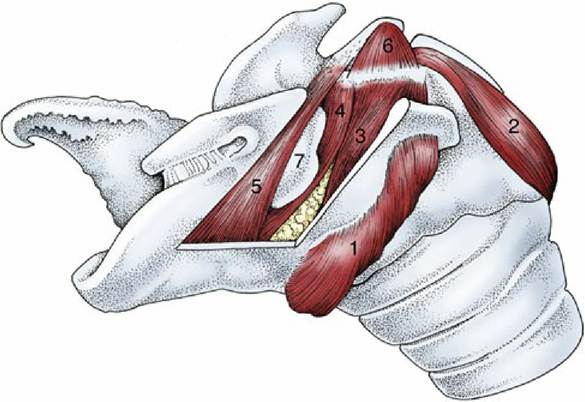
FIG. 4.12 Intrinsic muscles of the equine larynx. 1, Cricothyroideus; 2, cricoarytenoideus dorsalis; 3, cricoarytenoideus lateralis; 4, vocalis; 5, ventricularis (4 and 5 together: thyroarytenoideus); 6, arytenoideus transversus; 7, laryngeal ventricle.