The Cartilages
The forms of the laryngeal cartilages, and even the numbers of the minor elements, vary from species to species, but few differences are of great practical significance. The major, consistently present cartilages comprise the median epiglottic, thyroid, and cricoid cartilages and the paired arytenoid cartilages (Figs.
4.9 and 4.10).The epiglottic cartilage is most rostral. It consists of a small stalk and a large leaflike blade. The stalk is embedded between the root of the tongue, the basihyoid, and the body of the thyroid cartilage and is attached to all of these structures. At rest, the blade inclines dorsorostrally behind the soft palate (the retrovelar position), but it may be tilted backward to partially cover the entrance to the larynx when the animal swallows. It is composed of elastic cartilage and is flexible.
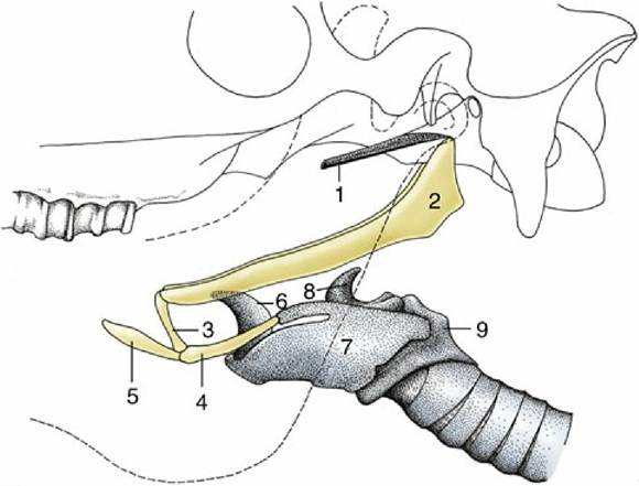
FIG. 4.8 Hyoid apparatus suspending the larynx from the base of the skull (horse). The broken line indicates the mandible. 1, Cartilage of auditory tube; 2, stylohyoid; 3, keratohyoid; 4, thyrohyoid; 5, lingual process of basihyoid; 6, epiglottic cartilage; 7, thyroid cartilage; 8, arytenoid cartilage; 9, cricoid cartilage.
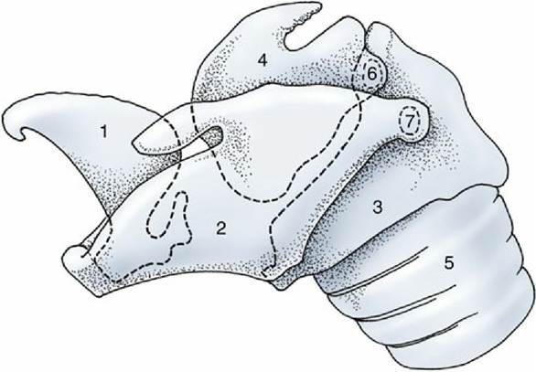
FIG. 4.9 Lateral view of the equine laryngeal skeleton. The outlines of those parts of the cartilages that are covered by others are indicated by broken lines. 1, Epiglottic cartilage; 2, thyroid cartilage; 3, cricoid cartilage; 4, arytenoid cartilage; 5, trachea; 6, cricoarytenoid joint; 7, cricothyroid joint.
The thyroid cartilage, the largest of the series, partly encloses the cricoid and arytenoid cartilages. It consists of two lateral plates that fuse to a varying degree ventrally and form a major part of the laryngeal floor (Fig.
4.10/3). The body formed by this ventral fusion is least extensive in the horse, in which a large, forward-pointing notch provides a convenient route of entry for laryngeal surgery. The most rostral part of the body is generally thickened and corresponds to the "Adam's apple," which is more salient in the human than in domestic species. The rostral and caudal extremities of the dorsal edge of each lamina articulate with the thyrohyoid cartilage and the arch of the cricoid cartilage, respectively. The thyroid cartilage is hyaline and so may become more brittle with age because of focal calcification and ossification.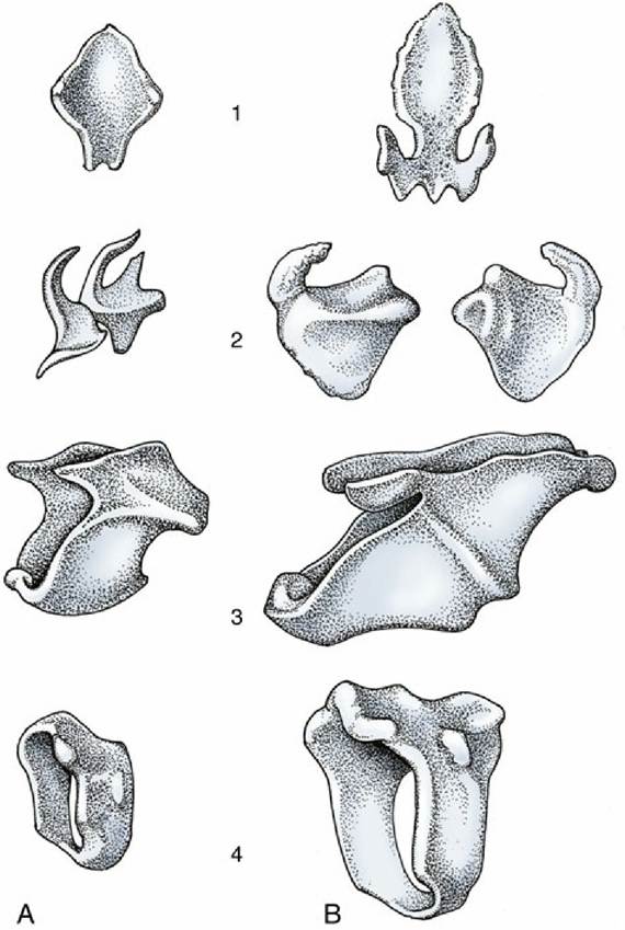
FIG. 4.10 Laryngeal cartilages of (A) the dog and (B) the horse. 1, Epiglottic cartilage; 2, arytenoid cartilage; 3, thyroid cartilage; 4, cricoid cartilage.
The cricoid cartilage is fashioned like a signet ring, consisting of an expanded dorsal "seal" (lamina) and a narrower ventral arch (Fig. 4.10/4). The dorsal part carries a median crest and, on its rostral rim, two facets for the arytenoid cartilages. The arch carries a facet on each side for articulation with the thyroid cartilage. The cricoid cartilage is also hyaline and subject to the aging process.
The arytenoid cartilages have a very irregular form best described as pyramidal (Fig. 4.10/2). The details are of little importance, so it is sufficient to recognize only a few features. A caudal facet articulates with the rostral margin of the cricoid lamina, and from it radiate (1) a vocal process that projects ventrally into the laryngeal lumen, and to which the vocal fold attaches; (2) a muscular process that extends laterally; and (3) a corniculate process that extends dorsomedially, forming the caudal margin of the laryngeal entrance with its fellow of the other side. The arytenoid cartilage is mainly hyaline, but the corniculate process is elastic.
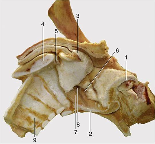
FIG. 4.11 Median section of the equine larynx after removal of the mucosa. 1, Epiglottic cartilage; 2, sectioned body of thyroid cartilage; 3, corniculate process of arytenoid cartilage; 4, sectioned lamina of cricoid cartilage; 5, cricoarytenoid joint; 6, ventricularis; 7, vocalis; 8, laryngeal ventricle; 9, tracheal rings.
Among the smaller and less prominent cartilages are the elastic cuneiform processes that support mucosal folds passing from the epiglottis to the arytenoids. These processes do not occur in all species, and when present, they may be free or fused with the epiglottis or with the arytenoid cartilages. A discrete nodule of hyaline cartilage, the interarytenoid cartilage, may be found between the arytenoid cartilages dorsally.