THE ARTICULATIONS OF THE JAWS
Although it is customary to describe two temporomandibular joints, these may be regarded as the widely separated halves of a single condylar joint (p. 21). Clearly, movement at one side must be accompanied by a movement, not necessarily identical, at the other side.
The articular surfaces are provided by the head, carried on a dorsal process of the ramus of the mandible, and the mandibular fossa of the skull, a facet mainly formed by the squamous temporal bone, although sometimes extending beyond it. The forms of the two surfaces reflect the feeding habits, and in species such as the dog, in which hingelike movements of the lower jaw predominate, the head takes the form of a transverse condyle to which the fossa provides a corresponding gutter. Backward dislocation of the jaw is opposed by the prominent retroarticular process placed directly behind the mandibular fossa. A peculiarity of the joint is the presence of a fibrous or fibrocartilaginous articular disk that divides the cavity into upper and lower compartments. Although the phylogenetic
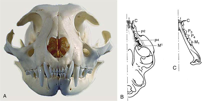
Figure 3-17 Permanent dentition of the cat. A, Rostral view. B, Upper jaw. C, Lower jaw.
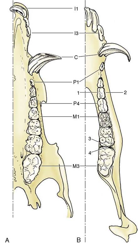
Figure 3-18 Permanent dentition of the pig, upper (A) and lower (B) jaws. 1, Lingual surface; 2, vestibular surface; 3, distal surface; 4, mesial surface.
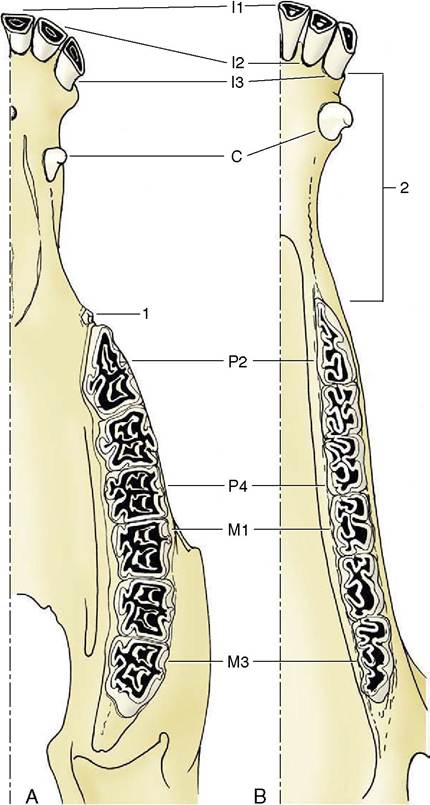
Figure 3-19 Permanent dentition of the horse, upper (A) and lower (B) jaws. 1, Wolf tooth (P1); 2, diastema.
origin of this structure is disputed, its functional significance may lie in its resolving the complex movements of the joint into simpler components; a hinge movement occurs between the mandible and the disk, while gross sliding movements (translations) of the mandible relative to the skull occur at the upper level.
It is perhaps because the movements of the dog’s jaw are so simple that the disk is rather thin and poorly developed in this species. In species in which lateral grinding movements predominate, the mandibular head is larger, the surface more plateau-like, and the disk thicker, although the details differ considerably.In most species the halves of the mandible are firmly fused together, but in the dog (and in ruminants) they articulate by means of a symphysis, providing a third joint. This much neglected joint allows small movements that may be important in securing more precise adjustment of the upper and lower tooth rows and therefore a more effective cutting or crushing mechanism. Two types of movement appear to be possible: a spreading movement, altering the angle between the halves of the mandible, and one in which each half rotates about its own long axis so that the tooth cusps alter their inclination to the vertical. The dog appears to make use of these possibilities when adjusting the position of a bone between the teeth before attempting to crack it.