THE MUSCLES OF MASTICATION
The muscles that provide the masticatory forces are derived from the first pharyngeal arch, and in keeping with this, they are supplied by the mandibular nerve.
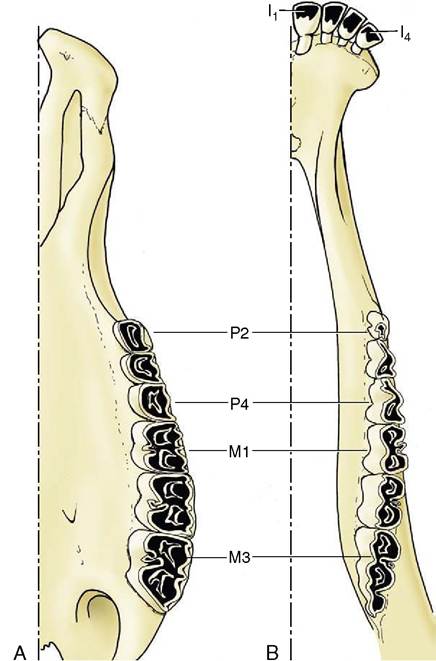
Figure 3-20 Permanent dentition of cattle, upper (A) and lower (B) jaws.
They comprise the temporalis, masseter, pterygoideus medialis, and pterygoideus lateralis (Figure 3-21). Other muscles that play some part in jaw movements, particularly in opening the mouth, are not normally included under the term muscles of mastication.
The temporalis arises from an extensive area on the lateral surface of the cranium and converges to an insertion on the coronoid process of the mandible. On contraction the resultant force pulls the mandible upward; the muscle is especially large in those species, such as the dog and cat, in which the chief jaw movement is scissorlike. A measure of its development is provided by the salience of the zygomatic arch: a well-sprung arch provides more room for this muscle. Although the main action is to raise the mandible, some fibers tend to draw it forward, while others tend to pull the condyle against the retroarticular process.
The masseter lies lateral to the mandible. It takes its origin from the maxillary region of the skull and the zygomatic arch and has a wide insertion on the more caudal part of the mandible. It is frequently a multipennate muscle intersected by strong tendon plates. The
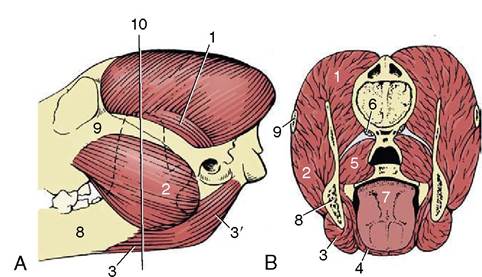
Figure 3-21 The muscles of mastication of the dog, left lateral aspect (A), in section (B). 1, Temporalis; 2, masseter; 3, 3', rostral and caudal bellies of digastricus; 4, mylohyoideus; 5, medial pterygoid; 6, origin of lateral pterygoid; 7, tongue; 8, mandible; 9, zygomatic arch; 10, level of transection (B).
fibers in the different strata do not all run parallel; different parts may have contrasting functions. Some may protrude the mandible, and others may retract it; however, the general effect is to raise the mandible and draw it toward the active side, for mastication is restricted to one side at a time in domestic species. The masseter muscle is therefore rather small in the dog; it is proportionately better developed in herbivorous species that make lateral and rotational movements when chewing.
The pterygoid mass of muscle lies medial to the mandible and passes to this bone from the pterygopalatine region of the skull. Generally the mass is clearly divided into a small lateral and a larger medial muscle. Some fibers of the lateral pterygoid muscle attach to the articular disk and help to control its movements, but the principal function of the mass is to raise the mandible and draw it inward with some simultaneous protrusion. In species in which transverse movements are important the masseter and contralateral pterygoid muscles may form a functional pair.
Opening the mouth is assisted by gravity, but certain muscles are also available for the performance of this movement. The digastricus passes from the skull, caudal to the temporomandibular joint, to the ventral margin of the mandible and opens the mouth. The muscle consists of two parts arranged in tandem. The rostral portion is supplied by the mandibular nerve, the caudal portion by the facial, which is an indication that the muscle has a composite origin in the mesoderm of the first two pharyngeal arches. In species in which the ster- nocephalicus has a mandibular attachment, it may open the mouth.
In most mammals the mouth is held closed at rest; the mandible is supported by the tonic activity of the masticatory muscles and possibly assisted by the hermetic seal created by the application of the dorsum of the tongue to the palate. The jaws are symmetrically placed in relation to the median plane, and the upper and lower tooth rows are slightly separated or in gentle, interrupted contact. The arcade formed by the upper teeth is generally wider than its counterpart, and the tooth rows are superimposed for only part of their widths.
In some species, such as the rat, simultaneous occlusion is impossible in both incisor and molar regions; in them, the lower jaw must be advanced and dropped to bring the incisor tips together and withdrawn and raised for molar contact. Such animals generally favor an intermediate position of the lower jaw at rest.A slight increase in muscular activity brings the teeth into more extensive contact, which is known as centric occlusion. The relationships between the teeth in this position are variable, even in the same individual at different ages since the teeth come together in altered fashion as wear reduces the more salient projections (and in some species also by migration of teeth within the jaws). It is usual to find that each cheek tooth engages with two teeth of the opposite series, and the lower teeth are generally a little mesial to their upper counterparts. In the dog, the largest teeth, the last upper premolar and the first lower molar, bite together and constitute the sectorial (or carnassial) teeth, the principal shear (see Figure 3-16). The teeth in front of the sectorials do not meet but leave open a carrying space, while the last cheek teeth make extensive contact. The lower canine engages in front of the upper canine, filling the space between this and the third incisor.
The relationship between the teeth is a dynamic one, as is readily seen from the so frequently defective human dentition. A tooth deprived of normal support may drift under the influence of the masticatory forces; the pressures exerted by the lips, cheek, and tongue are also important in maintaining normal contact and alignment. It is evident from developmental studies that these associations are established before eruption and that common factors control the growth of the two jaws and the development of the teeth so that a harmonious relationship normally exists at all stages of development. However, anomalies are not uncommon, and the “undershot” and “overshot” jaw are well illustrated by Bulldogs and by many Afghan Hounds.
The simplest activity that is common to all species, regardless of their masticatory habits, is the gaping that occurs on depression of the lower jaw. Gaping is achieved by slackening or cessation of activity in the masticatory muscles, by contraction of their antagonists, and by gravity. As the jaw is lowered the mandibular head rolls on the articular disk while the disk itself slides forward in the mandibular fossa, probably assisted by those lateral pterygoid fibers that attach to it. Closure of the mouth requires the reversal of these processes and must at times be vigorous enough to detach a morsel. Sometimes, the detachment is achieved by the incisors, and in certain species the hinge movement is complicated by a preliminary protrusion of the lower jaw to bring the incisor edges into alignment. When the cheek teeth are employed in biting, the action is unilateral. Herbivores employ the cheek teeth for grinding food already taken into the mouth, and the active (closing) movement is preceded by lateral displacement. The temporomandibular joint of these animals is situated high above the occlusal plane, and the lower teeth are drawn forward over their upper fellows as they approach. This contributes a grinding component that is absent when the joint and occlusal surfaces are more nearly level. The sheep and dog, typical examples of herbivore and carnivore, illustrate these differences in the position of the joint in relation to the teeth (Figure 3-22).