The auditory tubes open high on the lateral walls of the nasopharynx, immediately rostral to small mucosal cushions measuring about 10 mm long in dogs and 4 mm in cats.
Nasopharyngeal polyps, common in cats, originate in the middle ear as focal hypertrophies of the mucosa, develop stalks, and extend through the auditory tube to reach the nasopharynx.
There is a flat pharyngeal tonsil in the roof of the nasopharynx. Digital pressure in this area may stimulate respiration.The dorsoventrally flattened oropharynx extends from the palatoglossal arches, which stand out when the tongue is pulled forward. During normal breathing the soft palate lies on the tongue with its free edge rostral to the epiglottis (Figs. 11.29 and 11.33). In many brachycephalic dogs the soft palate is disproportionally long and rests over the entrance to the larynx, causing respiratory difficulties. The overlong soft palate can be shortened with the use of the palatine vessels laterally and the palatine muscle toward the midline as landmarks. Additional guidance is provided by the wrinkling of the palatine mucosa where it does not lie over muscle. For different reasons, the epihyoid provides a useful landmark where it crosses the lateral wall of the oropharynx. Contact with the oropharyngeal wall during examination of the mouth normally causes dogs to retch; the absence of this (gag) reflex suggests damage to the glossopharyngeal and vagal nerves.
Oral breathing is possible with the palate in the normal position (Fig. 11.30), and the panting dog is a familiar sight. Cats may also breathe through the mouth but more discretely, sitting quietly and letting the air slip in and out through lips slightly parted toward the commissure. Occasionally the mouth is opened more widely, allowing a brief glimpse of the tongue.
The fusiform palatine tonsils occupy fossae in the lateral walls of the oropharynx caudal to the palatoglossal arch and ventral to the soft palate and are covered medially by semilunar folds, which arise from the ventrolateral part of the soft palate (Figs.
11.17/8 and 11.31). In cats the palatine tonsil is very small and is covered by a mucosal fold.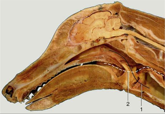
FIG. 11.30 Median section of head and neck. 1, Route from nasopharynx to trachea (solid line); 2, route of food from mouth to esophagus (broken line).
The tonsils are relatively large in young dogs and often protrude from the fossae; similar protrusion in the adult usually indicates pathologic swelling. In the performance of tonsillectomy the reddish lymphoid tissue that lines the fossa dorsal to the tonsil must also be removed; it is exposed when the main part is retracted from the fossa. The tonsil is related laterally to the lingual nerve and the mandibular and sublingual ducts, all of which are at some risk in this operation. The tonsil is supplied by tonsillar and hyoid branches of the lingual artery, which courses ventrolateral to the tonsil. Sensory innervation to the tonsil is from the glossopharyngeal nerve. The efferent lymph vessels drain to the medial retropharyngeal and mandibular lymph nodes. There are of course no afferents.
On each side the caudal border of the soft palate is continued to the dorsolateral wall of the