The Autonomic Nervous System Affects the Cardiovascular SystemThrough the Release of Epinephrine, Norepinephrine, and Acetylcholine
The autonomic nervous system is the “neuro” arm of neurohumoral control. Sympathetic and parasympathetic neurons influence the cardiovascular system through the release of the neurotransmitters norepinephrine and acetylcholine.
In addition, sympathetic nerves affect the cardiovascular system by stimulating the release of epinephrine and norepinephrine from the adrenal medulla. The adrenal secretions enter the bloodstream as hormones and circulate throughout the body. Chapter 13 contains additional, basic information about the autonomic nervous system.Whether acting as neurotransmitters or as hormones, epinephrine, norepinephrine, and acetylcholine exert their cardiovascular effects by activating membrane receptors on the cardiac muscle cells or on the endothelial cells or the smooth muscle cells of blood vessels. The receptors activated by epinephrine and norepinephrine are called adrenergic receptors (named after the adrenal gland). There are two major subtypes, alpha and beta: a-adrenergic receptors and β-adrenergic receptors. Each of these is subdivided into α∣, a2, β∣, and β2, and each of the four has important cardiovascular roles.
Acetylcholine activates cholinergic receptors. There are two major subtypes: muscarinic cholinergic receptors and nicotinic cholinergic receptors. The main cardiovascular effects of acetylcholine are mediated through muscarinic cholinergic receptors. Of five known types of muscarinic receptors, the M2 and M3 receptors have the greatest cardiovascular importance.
Table 25-1 summarizes the main cardiovascular consequences of the activation of adrenergic and cholinergic receptors. Alpha-adrenergic receptors (both α1 and α2) are located on the cell membranes of the smooth muscle cells of the arterioles in all organs of the body as well as on the smooth muscle cells of the abdominal veins.
The α-adrenergic receptors on arterioles and abdominal veins are innervated by postganglionic sympathetic neurons. Activation of the α-adrenergic receptors leads to constriction of the arterioles or the veins.Arteriolar vasoconstriction increases the resistance and decreases the blood flow through an organ. If one or more major body organs are Vasoconstricted, the TPR increases. TPR (along with cardiac output) determines arterial blood pressure, so widespread α-adrenergic vasoconstriction in the body leads to an increase in arterial blood pressure. The increase in arterial pressure increases the driving force for blood flow in the nonvasoconstricted organs. In this way, the sympathetic nervous system can use vasoconstriction in some organs to direct more blood flow to other, nonvasoconstricted organs.
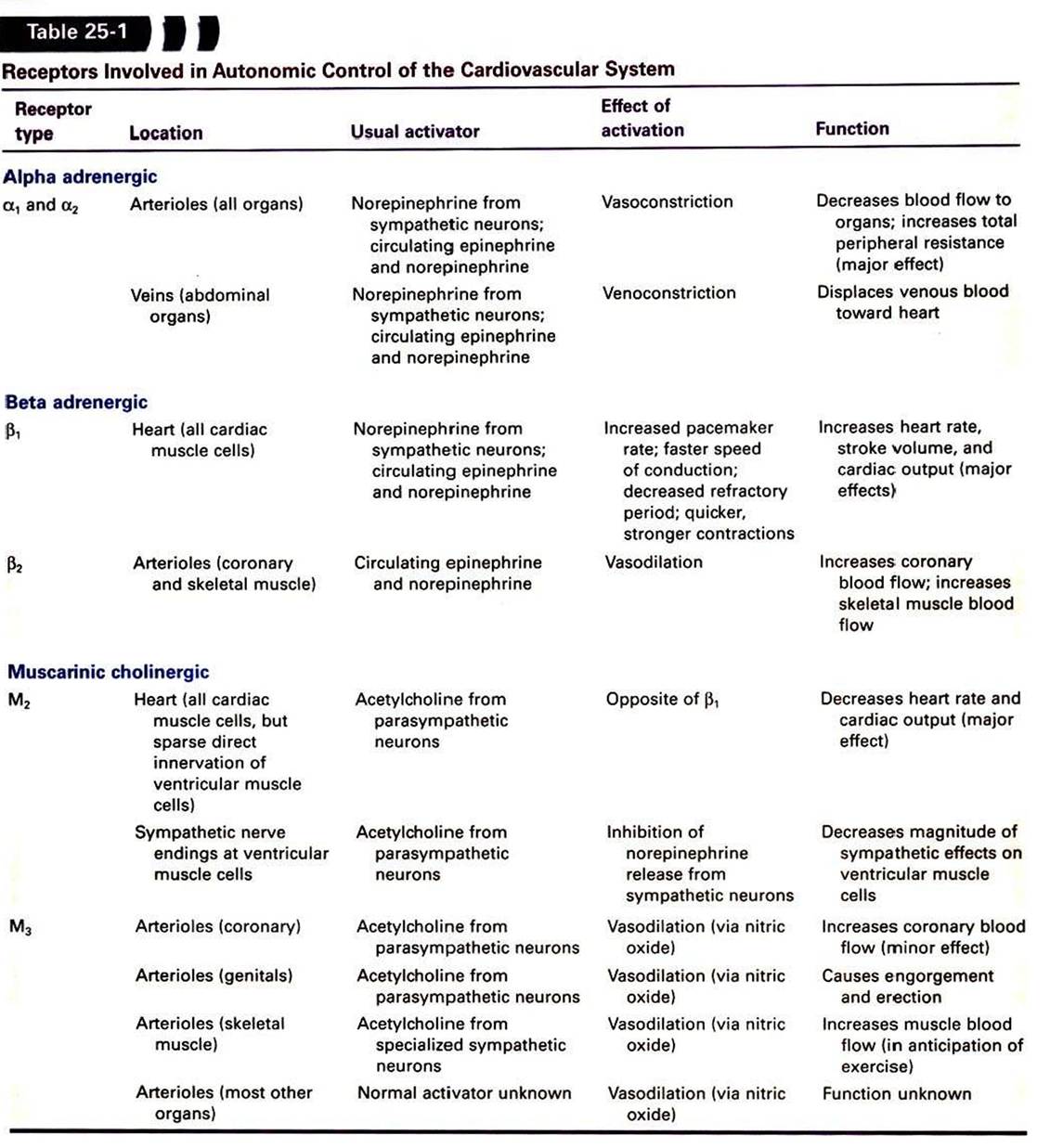
The major role of veins is to act as reservoirs for blood. Venoconstriction displaces venous blood toward the central circulation, which increases central venous pressure, ventricular preload, and stroke volume. Venoconstriction in the abdominal organs is particularly important in the control of central blood volume and therefore central venous pressure. Venoconstriction causes only a small increase in the resistance to blood flow through an organ because the veins, whether constricted or dilated, offer much less resistance to blood flow than do the arterioles.
Sympathetic control of the heart is exerted through the β∣- adrenergic receptors, which are found on every cardiac muscle cell. These β-adrenergic receptors are activated by norepinephrine or epinephrine. Chapters 19 and 21 discuss the effects of activation of the cardiac β-adrenergic receptors. In brief, pacemaker rate increases, cell-to-cell conduction velocity increases, and refractory period decreases. In addition, contractility is increased, so the cardiac contractions are quicker and stronger.
The overall effect is increased heart rate and stroke volume.The β>-adrenergic receptors are found on the arterioles, particularly in the coronary circulation and in skeletal muscles. The activation of arteriolar β2-adrenergic receptors causes relaxation of the vascular smooth muscle and dilation of the arterioles. However, these β2-adrenergic receptors are not innervated by the sympathetic nervous system, so they are not activated directly by sympathetic nerves. Instead, they respond to circulating epinephrine and norepinephrine (released from the adrenal medulla). The adrenal medulla releases epinephrine and norepinephrine in situations that involve trauma, fear, or anxiety. Dilation of arterioles in the coronary circulation and in skeletal muscles is appropriate in such “fear, fight, or flight” situations because the dilation results in an anticipatory increase in blood flow to the heart and skeletal muscle. /Xppropriately for its role in emergency situations, β2-adrenergic vasodilation can overpower α- adrenergic vasoconstriction in the coronary circulation and in skeletal muscles.
Cholinergic muscarinic receptors of the M2 type are located in the cell membranes of cardiac muscle cells. The natural stimulus for the cardiac M2 receptors is acetylcholine, released from postganglionic parasympathetic neurons. Cells of the sinoatrial and atrioventricular nodes are densely innervated by postganglionic parasympathetic neurons. Atrial cells also receive strong parasympathetic innervation. In the sinoatrial node, atria, and atrioventricular node, the activation of M2 receptors has effects basically opposite to those of the activation of β∣-adrenergic receptors. Parasympathetic activation powerfully slows the pacemaker rate, decreases the cell-to-cell conduction velocity, and increases the refractory period. By contrast, few ventricular muscle cells receive direct parasympathetic innervation.
Therefore, parasympathetic activation has very minor, direct effects on cardiac contractility. However, parasympathetic activation can still profoundly decrease cardiac output by decreasing the heart rate. In addition, parasympathetic neurons do exert an interesting, indirect effect on ventricular muscle cells. Most parasympathetic neurons in the ventricles release their acetylcholine onto sympathetic neuron terminals, rather than directly onto ventricular muscle cells. This acetylcholine activates muscarinic cholinergic receptors on the sympathetic neuron terminals, which inhibits the release of norepinephrine from the terminals and thus weakens the effects of sympathetic activity on ventricular cells.Cholinergic muscarinic receptors of the M5 type arc found on the endothelial cells and on the smooth muscle cells of most arteries and arterioles. Activation of M5 receptors on smooth muscle cells causes them to contract (and the blood vessels to constrict). However, this vasoconstrictor effect is usually overridden by the vasodilatory effect of activating the M5 receptors on the vascular endothelial cells. In this strange arrangement, activation of M5 receptors on endothelial cells causes the synthesis of nitric oxide, which then diffuses out of the endothelial cells and into the nearby smooth muscle cells, where it causes vasodilation. The vasodilatory effect of stimulating the M3 receptors on endothelial cells is stronger than the vasoconstrictor effect of stimulating the M5 receptors on smooth muscle cells.
The M5 receptors on vascular endothelial cells are innervated in three tissues. In the coronary circulation, parasympathetic neurons innervate vascular M5 receptors and bring about vasodilation. This vasodilatory effect is minor, however, and the function of this innervation is unclear. In the external genital organs, parasympathetic neurons release both acetylcholine and nitric oxide.
The acetylcholine acts on M5 receptors to stimulate the release of additional nitric oxide from endothelial cells. The nitric oxide causes vasodilation, engorgement of the organs with blood, and erection. The third tissue in which vascular M3 receptors are innervated is skeletal muscle. In some species (e.g., cats and dogs) but not in others (e.g., primates), the M3 receptors of skeletal muscle blood vessels are innervated by special postganglionic sympathetic neurons that release acetylcholine (rather than norepinephrine) as a neurotransmitter. These sympathetic cholinergic neurons appear to be activated specifically in anticipation of muscular exercise and during the “fear, fight, or flight” (defense-alarm) reaction. The resulting vasodilation increases blood flow through the skeletal muscle just before and during the initiation of exercise. Although primates do not have sympathetic cholinergic vasodilatory nerves, they can bring about an anticipatory vasodilation of skeletal muscle arterioles in another way, through activation of β-adrenergic receptors by circulating epinephrine and norepinephrine, as mentioned earlier.To summarize, although arteries and arterioles throughout the body dilate when exposed to acetylcholine, only the arterioles of the heart, external genitalia, and (in some species) skeletal muscle are innervated by acetylcholine-releasing autonomic neurons. The functional significance of the M3 receptors on arterioles in other organs is unclear because no neurons (either sympathetic or parasympathetic) appear to innervate them, and neither acetylcholine nor any other muscarinic receptor agonist normally circulates in the bloodstream.
Of all the autonomic influences on the cardiovascular system just mentioned, three stand out as most important. The first is α∣- and α2-adrenergic vasoconstriction in the arterioles of all body organs, which is brought about by the sympathetic nervous system. The second is β1-adrenergic excitation of cardiac muscle, which is brought about by the sympathetic nervous system and results in an increased heart rate and stroke volume. The third is the decrease in heart rate brought about by activation of cardiac M2 receptors.
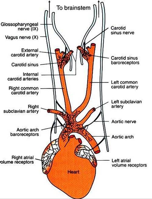
FIGURE 25-1 ■ Arterial baroreceptors are located in the walls of the carotid sinuses and in the walls of the aortic arch and its major branches.The atrial volume receptors are located in the walls of the right and left atria. See text for a description of the neural pathways followed by the baroreceptor and volume receptor afferents.