The Blood Flow to Each Organ Is Determined by the Perfusion Pressure and the Vascular Resistance
If the equation that defines resistance is solved for flow, the result is:
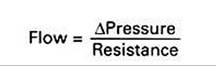
As applied to the blood flow through any organ, this equation points out that the blood flow is determined by the perfusion pressure (arterial pressure minus venous pressure) and by the resistance of the blood vessels of the organ.
There are no other factors. AU the organs of the systemic circulation are exposed to the same perfusion pressure. Therefore the differences in blood flow to the various organs result solely from their different vascular resistances. As explained earlier, the vascular resistance of an organ is determined primarily by the diameter of its arterioles. Thus, arteriolar vasodilation and vasoconstriction are the primary mechanisms that increase or decrease the blood flow in one organ relative to another organ.This point is illustrated in Figure 22-6. In a typical dog at rest, the arteriolar resistances are similar (diameters are the same) in the splanchnic, renal, and skeletal vascular beds. Therefore, each of these beds receives about the same blood flow (shown in Figure 22-6 by arrows of equal width). During exercise, skeletal muscle arterioles dilate greatly, almost doubling in diameter, which decreases their resistance to blood flow by a factor of almost 16. Therefore the skeletal muscle blood flow increases almost 16-fold (from 0.5 to 7.8 L∕min). Also during exercise, coronary arterioles dilate, so the coronary blood flow increases. Brain arterioles remain the same, so the brain blood flow is unchanged. By contrast, the arterioles in the splanchnic and renal circulations constrict slightly during exercise, which causes splanchnic and renal resistance to increase by about 20%. Therefore the splanchnic and renal blood flows decrease by about 20% (from 0.5 to 0.4 L∕min).
This discussion of blood flow during exercise describes the responses of a normal dog with a healthy heart. Such a dog can readily increase its cardiac output enough to meet the increased blood flow needs of the skeletal and cardiac muscle. As a consequence, the arterial pressure and the perfusion pressure are very similar during rest and exercise. By contrast, a dog with heart failure cannot increase the cardiac output much above its resting level. Therefore the arterial pressure (and perfusion pressure) declines during exercise, and none of the organs receives the blood flow that it requires. This is why animals with heart failure exhibit weakness, fatigue, and exercise intolerance. (Additional complications of heart failure are discussed in Chapter 26.) The point for now is that the equation that relates blood flow, perfusion pressure, and vascular resistance is fundamental and inescapable; this relationship is profoundly important to an understanding of cardiovascular function and dysfunction.