The Pulmonary Circulation Offers Much Less Resistance to Blood Flow Than Does the Systemic Circulation
As with any other resistance, pulmonary resistance is calculated as a pressure difference (perfusion pressure) divided by a flow. The perfusion pressure that forces blood through the pulmonary circuit is the pressure in the pulmonary artery minus the pressure in the pulmonary veins.
The flow that traverses the pulmonary circuit is equal to the cardiac output. Therefore:
For a typical dog at rest, the mean pulmonary arterial pressure is 13 mm Hg, the mean pulmonary venous pressure is 5 mm Hg, and the cardiac output is 2.5 L∕min. Thus, pulmonary resistance is 3.2 mm Hg/L/min. Note that this is only about one- twelfth the resistance of the systemic circulation.
The entire cardiac output passes through the lungs, so a fourfold increase in cardiac output during exercise also necessitates a fourfold increase in pulmonary blood flow. Pulmonary blood vessels are quite compliant, and they readily distend to accept the increase in blood flow. Because even a small increase in vessel radius greatly decreases resistance (in accordance with PoiseuilIes law, as mentioned earlier), the resistance of the pulmonary blood vessels drops greatly during exercise. The decreased pulmonary resistance during exercise is advantageous because it allows the pulmonary flow to increase greatly without necessitating a large increase in the pulmonary arterial pressure.
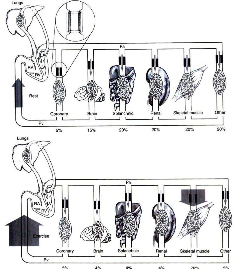
FIGURE 22-6 Cardiac output and its distribution compared during rest (top) and vigorous exercise (bottom) in a typical large dog.The width of the shaded arrows denotes the amount of blood flow.The flow of blood into the right side of the heart (which is equal to the cardiac output) is represented by the very wide arrows on the left.
The cardiac output is 2.5 Umin at rest and increases to 10.0 Umin during exercise (fourfold increase).The entire cardiac output passes through the lungs and then is pumped by the left ventricle (LV) into the systemic arterial system (horizontal tube across top).The systemic arteries deliver blood to each of the systemic vascular beds, which are grouped here into coronary, brain, splanchnic, renal, skeletal muscle, and other. In each systemic organ, blood must pass through high-resistance arterioles (heavy bars) before reaching the capillaries.The arterioles act as adjustable cuffs or constrictors (see magnified view, top).The proportion of the total cardiac output that goes to each organ is indicated by a percentage at the bottom. Because each organ is exposed to the same arterial pressure (Pa) and venous pressure (Pv)t the proportion of cardiac output that each organ receives is determined by its resistance. Resistance is determined primarily by the arteriolar diameter, which is indicated by the size of the opening between the heavy bars. During vigorous exercise, skeletal muscle arterioles dilate maximally, and the blood flow to the exercising muscles increases 16-fold (from 0.5 Umin at rest to 7.8 Umin). Coronary arterioles also dilate, and the coronary blood flow increases about fourfold, which meets the increased demand by the heart muscle for oxygen. Vasoconstriction causes a small decrease in blood flow to the splanchnic and renal circulations. Blood flow to the brain is basically unchanged. Of course, the percentage of total cardiac output received by the brain decreases. RVt Right ventricle; LAt left atrium; RAt right atrium.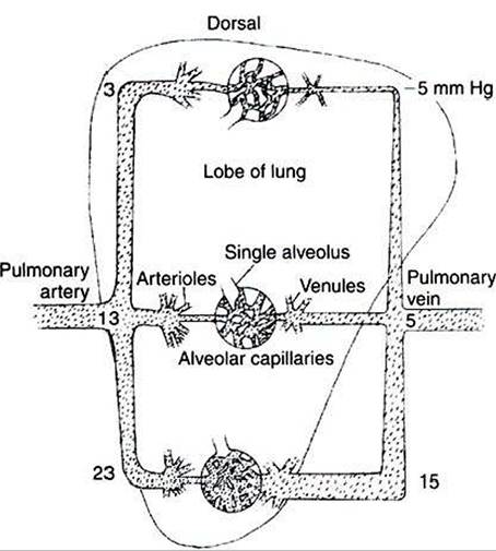
FIGURE 22-7 Gravity pulls downward on the blood within the lungs, which increases the pressure within blood vessels low in the lungs (see mm Hg values in the figure).The pressure outside the blood vessels (intrapleural pressure) is not affected much by gravity (because air is so much lighter than blood).Therefore the low-lying blood vessels become distended, which decreases their resistance to blood flow. As a result, more blood flows through the lower parts of the lungs than through the upper parts.
In other circumstances the high compliance of pulmonary vessels can lead to adverse consequences. An example is the effect of gravity on pulmonary blood flow. Gravity pulls downward on the blood within lung blood vessels, which increases the blood pressure in vessels low in the lungs compared with vessels that are higher (Figure 22-7). The greater pressure distends the vessels low in the lungs, which decreases their resistance to blood flow. As a consequence, more of the pulmonary blood flow traverses the lower (dependent) regions of a lung than the higher regions. For reasons explained later (see Chapter 46), gravity also causes more air to be delivered to the dependent regions of the lungs than to the higher regions. However, gravity has a greater effect on blood flow than on air delivery, so blood flow tends to be excessive (relative to air delivery) in the dependent region of a lung. Any such imbalance in the lungs between air delivery and blood flow is called a ventilation-perfusion mismatch. This inherent problem is most severe in large animals, in which the large size of the lungs leads to substantial gravitational effects.
Hypoxic vasoconstriction is an important mechanism that helps offset ventilation-perfusion mismatches in the lungs regardless of whether these mismatches result from gravitational effects or from any other cause. The pulmonary blood vessels are sensitive to the local concentration of oxygen (measured as oxygen partial pressure, Po2). A low Po2 (hypoxia) causes pulmonary vessels to constrict. Hypoxic vasoconstriction takes place in any region of the lung where ventilation (the
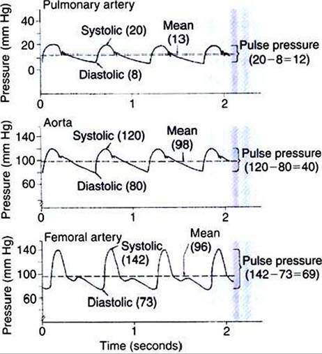
FIGURE 22-8 Blood pressure in the large arteries is pulsatile.The pressure patterns typical of the pulmonary artery, aorta, and femoral artery of the dog are shown.
delivery of fresh air and oxygen) is reduced relative to blood flow. The vasoconstriction increases the resistance of blood vessels in that lung region and thereby reduces blood flow (perfusion).
In this way, hypoxic vasoconstriction brings about a better match between ventilation and perfusion.As with many compensatory mechanisms, hypoxic vasoconstriction can have undesirable consequences. For example, consider what happens if ventilation becomes depressed throughout the lungs. This may occur during an allergic constriction of the airways (asthma) or as the result of chronic obstructive pulmonary disease, which is common in horses. Depressed ventilation causes hypoxic vasoconstriction throughout the lungs. Pulmonary vascular resistance increases greatly, which necessitates a substantial increase in pulmonary arterial pressure to maintain pulmonary blood flow. The condition of elevated pulmonary arterial pressure is called pulmonary hypertension. Pulmonary hypertension greatly increases the workload of the right ventricle. In extreme cases, it leads to right ventricular failure.