THE BLOOD SUPPLY OF THE EYE
The blood supply to the eyeball and its adnexa is complex (Figure 9-22). The blood supply to the human eye enters the orbit with the optic nerve. This route is represented in the domestic mammals by the rudimentary internal ophthalmic artery (Figure 9-22/2), which loses its identity when joined by a sizable anastomosis (Figure 9-22/4) from the external ophthalmic.
The principal supply is carried by the external ophthalmic artery (Figure 9-22/5), a branch detached from the maxillary as this passes ventral to the orbit to supply more rostral structures of the face. The arteries arising from the external ophthalmic and malar arteries (a further, smaller branch of the maxillary) can be divided into three groups: (1) those supplying the eyeball, (2) those supplying ocular muscles, and (3) those leaving the orbit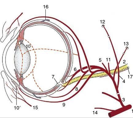
Figure 9-22 The principal arteries supplying the eye. 1, Maxillary a.; 2, rudimentary internal ophthalmic a.; 3, external ophthalmic a.; 4, anastomosis between external and internal ophthalmic aa.; 5, lacrimal a. to lacrimal gland and upper lid; 6, short posterior ciliary aa.; 7, retinal aa.; 8, long posterior ciliary aa.; 9, anterior ciliary aa., substantial branches to 10 in horse, lesser branches in the other domestic species; 10, greater arterial circle of the iris; 10', annular pericorneal network; 11, muscular branches; 12, supraorbital a. and foramen; 13, external ethmoidal a. and foramen; 14, malar a.; 15, palpebral branches; 16, vorticose veins; 17, optic nerve.
to supply adjacent structures, regardless of whether these are associated with the eye.
1. The branches of the external ophthalmic artery for the eyeball penetrate the sclera to reach the vascular tunic and the retina. Short posterior ciliary arteries (Figure 9-22/6) penetrate near the optic nerve and supply the adjacent choroid in addition to branches to the optic nerve.
The latter form the central artery of the retina, the parent vessel for the retinal arteries (Figure 9-22/7; Figure 9-6, A-F). Long posterior ciliary arteries (Figure 9-22/8) pass through the sclera somewhat closer to the equator. The anterior ciliary arteries (Figure 9-22/9) penetrate near the limbus and supply the anterior portion of the choroid, the ciliary body, and the iris. These arteries anastomose to form the greater arterial circle of the iris (Figure 9-22/10) from which numerous fine branches pass toward the pupil and into the ciliary body. Capillaries near the limbus nourish the cornea by diffusion. The anterior ciliary arteries also send branches to the conjunctiva (Figure 9-23). The principal venous return is by several vorticose veins (Figure 9-22/16) that emerge from the sclera near the equator. The extraocular veins of carnivores and ruminants form substantial venous plexuses within the periorbita. Venous blood returning from the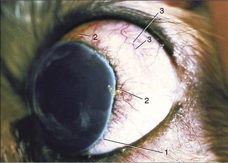
Figure 9-23 Exophthalmic canine eyeball and associated vascularization of bulbar conjunctiva and anterior sclera.
retina leaves at the optic disc through small veins satellite to the short posterior ciliary arteries.
2. No more need be said about the arteries supplying the ocular muscles except that most enter the muscles proximally. The absence of larger vessels in the distal ends reduces bleeding when the muscles are cut during enucleation.
3. Only four of the arteries that leave the orbit require mention. The lacrimal artery (Figure 9-22/5) passes forward in the lateral part of the orbital cone and, after supplying the lacrimal gland en route, crosses the dorsolateral part of the orbital margin to supply lateral parts of the eyelids and conjunctiva. The supraorbital artery (Figure 9-22/12) passes dorsally and leaves the orbit by the supraorbital foramen. It ramifies subcutaneously medial to the orbit and may send branches into the upper eyelid.
(Carnivores have no supraorbital foramen and artery; the blood supply to their eyelids comes from long branches of the superficial temporal artery.) The malar artery (Figure 9-22/14) arises directly from the maxillary and passes over the ventral wall of the orbit to the medial angle of the eye, where it supplies the eyelids and also the adjacent area of the face. The external ethmoidal artery (Figure 9-22/15) has the shortest intraorbital course of the four. It leaves the orbit through the ethmoidal foramen and supplies the ethmoid labyrinth of the nasal cavity.Most of the arteries described also take part in supplying the fat, fascia, and nerves within the orbit. There is some interspecific variation, but this is rarely of practical concern. However, it may be noted that the external ophthalmic artery of the ruminants breaks up and forms a small arterial network (rete mirabile ophthalmicum) on entering the orbit. The various arteries, except the malar, arise from this.