» The Carpus and Forepaw (see also pp. 71-75 and 80)
The carpal and metacarpal bones and the phalanges should be studied principally with a view to becoming familiar with their radiographic appearances.
The most obvious external features are the digital, metacarpal, and carpal pads and the claws.
At birth, a reduced first digit, or "dewclaw," is generally present below the carpus on the medial side of the paw. It is often removed routinely, even in city dogs, although the presumed purpose of this mutilation is to avoid the risk of injury should the dewclaws catch in scrub. It must be retained in puppies of certain breeds if there is a possibility that they will later be shown. The carpal pad, just distal to the palpable accessory carpal bone, is normally denied contact with the ground except in animals cornering at speed; it is occasionally injured in this way in racing Greyhounds (see Fig. 10.15/4). The metacarpal (Fig. 16.9/8) and digital (Fig. 16.9/7) pads over flexor surfaces of the metacarpophalangeal and the distal interphalangeal joints, respectively, make ground contact, and the small papillae that normally roughen their surfaces may be worn smooth in dogs regularly walked on pavement. The webs of skin connecting the digits proximal to the pads are common sites of interdigital infections and cysts.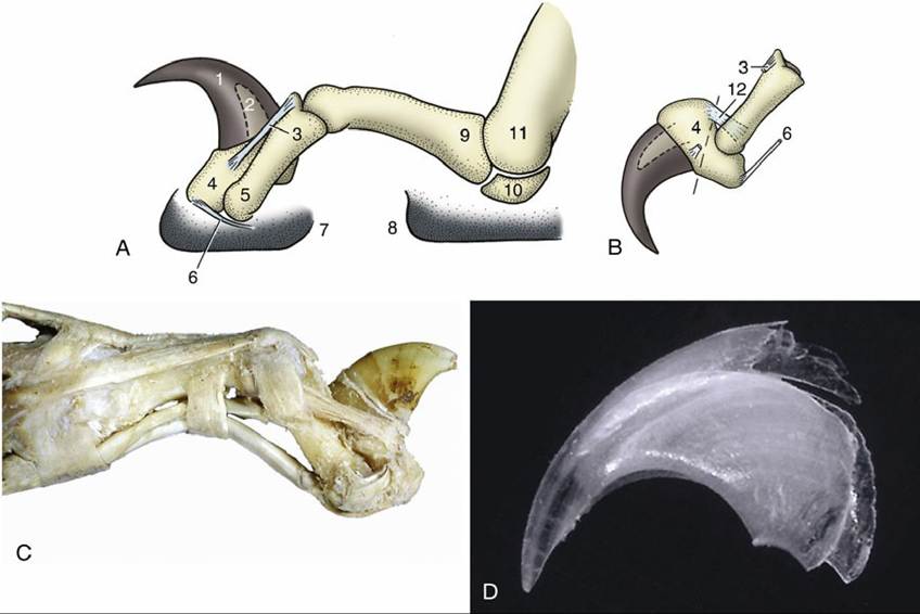
FIG. 16.9 The feline claw (A) fully retracted and (B) protruded, showing the division (broken line) of the distal phalanx in declawing. The arrangement of the elastic ligaments has been greatly simplified. (C) Claw of a tiger showing the same ligaments somewhat more clearly. (D) Outer layer of horn shed from cat claw. 1, Claw; 2, unguicular process of distal phalanx; 3, medial dorsal elastic ligament; 4, distal phalanx; 5, middle phalanx; 6, deep digital flexor tendon; 7, digital pad; 8, metacarpal pad; 9, proximal phalanx; 10, proximal sesamoid bone; 11, metacarpal bone; 12, lateral dorsal elastic ligament.
Dorsopalmar radiographs show the carpal bones with a minimum of overlapping (Fig. 16.10). The large radial carpal bone (Fig. 16.10/3), which incorporates the intermediate element in both dogs and cats, lies distal to the radius; the oddly shaped ulnar carpal bone (Fig. 16.10/4) next to it extends distally (on the palmar surface) to be superimposed on the fourth carpal bone (and even on the corresponding metacarpal bones). The accessory carpal bone (Fig. 16.10/5) is superimposed on the junction of the radius, ulna, and ulnar carpal bone. On the medial side, carpals 1 and 2 are superimposed, and a sesamoid in the extensor carpi obliquus may also be seen opposite the midcarpal joint. Another two sesamoid bones may be visible on the palmar aspect between the proximal and distal carpal rows. The carpal pad produces a fainter shadow. The distal radial epiphysis has occasionally been mistaken for a carpal bone. A wide space seen between the distal extremities of the radius and ulna in slightly oblique projections of the cat's carpus may be misinterpreted as a subluxation.
The proximal row of carpal bones includes the fused radial, intermediate, and central bone, the ulnar carpal bone, and the accessory carpal bone (see Fig. 2.48). The radial carpal bone exhibits three ossification centers that fuse 3 to 4 months after birth in dogs, although not until the seventh month in the cat. Although the ulnar carpal bone has a large distally protruding process, it possesses only a single ossification center. The epiphysis of the accessory carpal bone closes between 3 and 6 months of age. The distal carpal row is composed of four bones, the smallest of which is medial and the largest of which is lateral.
The antebrachiocarpal joint is an ellipsoid joint allowing flexion, extension, abduction, and adduction. In dogs and cats the collateral ligaments do not extend the length of the carpus but are limited to the proximal joint. Short carpal ligaments bridge the chief joints vertically, connect neighboring bones in the same row horizontally, and connect the accessory carpal bone to the ulna, the ulnar and fourth carpal bones, and the fourth and fifth metacarpal bones.
Only the two distal joint spaces communicate, while the independent proximal (antebrachiocarpal) compartment may be punctured most readily by passing the needle between the palpable radial carpal and common digital extensor tendons when the joint is flexed. Flexion of the joint widens the dorsal gap at the antebrachiocarpal level and facilitates appreciation of the tendons of the extensor carpi radialis and common digital extensor. Except for the accessory, the individual carpal bones cannot be distinguished by palpation. The bones distal to the carpus are all readily identified by palpation because the metacarpals, though crowded together proximally, diverge distally. The extensor tendons can be rolled against the metacarpal bones, and the digital flexors and the interossei together form a soft package on the palmar aspect.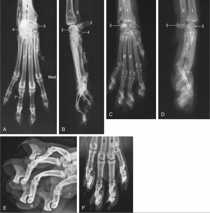
FIG. 16.10 (A to D) Dorsopalmar and lateral radiographic views of the canine (A and B) and feline (C and D) forepaws. (E) and (F) Oblique and dorsopalmar views of feline digits, respectively; note how the distal phalanges slide next to the middle phalanges when the claws are retracted. 1, Radius; 2, ulna; 3, radial carpal; 4, ulnar carpal; 5, accessory carpal; 6 and 6', third and fourth metacarpals, respectively; 7, metacarpal pad; 7', distal border of metacarpal pad, 8, digital pad; Med., medial.
The distal epiphyses of the principal metacarpal bones fuse with the shafts at about 5 to 7 months. (The proximal metacarpal epiphyses fuse prenatally.)
The paired sesamoid bones on the palmar surface of the metacarpophalangeal joints are embedded in the metacarpal pad (Fig. 16.9/8 and 10). The sesamoid bones at the metacarpophalangeal joint are associated with the same complex of ligaments—straight, oblique, and so forth—as in the horse but without these possessing corresponding importance. Distal to the proximal sesamoid bones, the branches of the superficial tendon are split for the passage of the deep tendon, and at the metacarpophalangeal and proximal and distal interphalangeal joints, these are retained by annular ligaments.
The functional digits (numbers 2 to 5) are equipped with interosseous muscles on the palmar aspects of the metacarpal bones, where their presence may be appreciated on deep palpation. In addition, digits 1, 2, and 5 each contain several small individual muscles of restricted functional and minimal clinical importance.The claws are shaped to the dorsal and lateral surfaces of the curved unguicular processes of the distal phalanges to which they are connected by the laminar dermis (Fig. 16.11B and C). The sole of each claw (Fig. 16.11/4) covers the ventral surface of the process and appears as a crumbly whitish material between the lower edges of the wall. The claws, especially those of heavy city dogs, are generally worn level with the digital pads; they must be trimmed when there is insufficient wear because, if left unchecked, they would grow around to penetrate the pads. Special clippers should be used because the lateral pressure exerted by scissors or human nail clippers causes pain. The claw should be trimmed level with the ground surface of the pad but not so short that the vascular and sensitive dermis is damaged (Fig. 16.11B). The pink dermis may be recognized in nonpigmented claws, but when one is denied this guide, a warning sign is provided by the appearance of a black dot on the cut surface just distal to the dermis.
Elastic dorsal ligaments (Fig. 16.11/5) extend from the proximal ends of the middle phalanges to the unguicular crests of the distal phalanges to keep the claws elevated. The deep digital flexor opposes the ligaments and protrudes the claws for scratching or digging.
The claws of the cat are laterally compressed, strongly curved, and drawn out to sharp points. They can be fully retracted into the fur of the paw, which enables cats to walk silently and without blunting the claws through ground contact. The elastic dorsal ligaments are of unequal length; long ones extend from the proximal interphalangeal joint to the sides of the distal phalanx, and a single short ligament extends between the distal end of the middle phalanx and the top of the unguicular crest (Fig.
16.9/3 and 12). This disposition, combined with the obliquity of the articular surfaces, allows the base of the claw to be drawn lateral to the corresponding middle phalanx (Fig. 16.10F).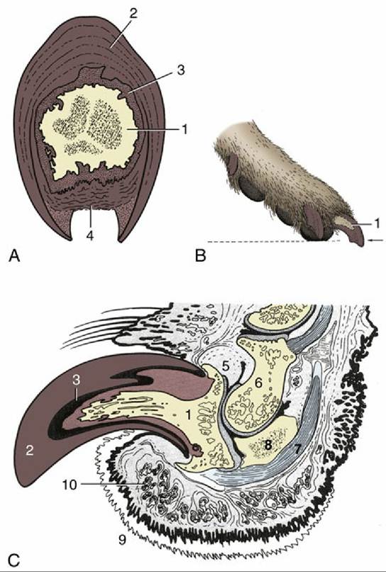
FIG. 16.11 (A) Transverse section of canine claw. (B) Correct trimming of canine claws. (C) Axial section of the canine digit. 1, Unguicular process of distal phalanx; 2, wall of claw; 3, laminar dermis; 4, crumbly sole of claw; 5, dorsal elastic ligament; 6, middle phalanx; 7, deep digital flexor tendon; 8, distal sesamoid (cartilaginous); 9, digital pad; 10, sweat glands.
The ligaments keep the claws strongly retracted so that the digital flexors move only the metacarpophalangeal and proximal interphalangeal joints. The claws are protruded by simultaneous contraction of the deep digital flexor, which flexes the distal interphalangeal joints, and the digital extensors, which stabilize the more proximal joints of the paw. Cats use their protrusible claws for climbing trees and for initial prey contact; dogs, however, use their jaws for prey contact. The characteristic "clawing" of cats on logs, rugs, or furniture, commonly thought to be performed to sharpen the claws, is actually related to territorial marking by sweat from the glands concentrated in the digital pads. Forceful scraping of the ground by dogs after defecation or urination may have a similar marking purpose that utilizes the secretion of the sweat glands of their pads. Clawing also promotes shedding of an outer, worn-out layer of a claw (Fig. 16.9D).
Onychectomy is the surgical procedure to remove P3 (the third digital phalanx) in destructive cats to prevent them from scratching furniture or people or to remove an infected nail or a nail tumor. The elective procedure is typically done between 3 and 12 months. The base of the bone with the attachment of the deep digital flexor is left in place while the unguicular crest, enclosing the base of the claw, is removed (Fig. 16.9B). The surgery is performed under general anesthesia and with nerve blocks. An alternative procedure, simpler and causing less postoperative pain, consists of deep digital flexor tenectomy. These procedures are forbidden in many European countries.
The main arteries of the forelimb have been described (pp. 231-232); their relations are shown in Fig. 16.5. A branch of the radial artery found on the dorsomedial aspect of the distal carpus may be used for taking the pulse of cats.