THE CAUDAL PART OF THE FOREGUT
A fusiform enlargement identifies the stomach at an early stage. The foregut between this and the pharynx becomes the esophagus, which is initially very short but elongates as the heart descends from the neck into the thorax.
The esophagus is involved in the origin of the lower respiratory tract (p. 165) but, apart from this, presents little of interest. At one stage, the proliferation of the endodermal lining obstructs the lumen, but the passage is later restored.The development of the stomach involves displacement, reorientation, and differential enlargement. The displacement carries it to a position ventral to the caudal thoracic segments. Reorientation appears to involve rotations about two axes. Rotation about the long axis of the stomach spindle carries the originally dorsal aspect to the left, where it is later distinguished as the greater curvature. The dorsal megogastrium, which becomes the greater omentum, shares in the process. Rotation about a vertical axis swings the cranial (cardiac) extremity to the left and the caudal (pyloric) one to the right (Figure 3-61). In most species the most conspicuous change in shape is an asymmetrical enlargement to the left of the cardia that produces the fundus; a much more radical reshaping is required in ruminants.
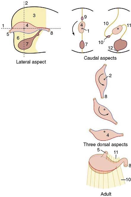
Figure 3-61 The reorientation of the developing simple stomach. It rotates counterclockwise (as seen from behind) around a longitudinal axis (caudal aspects [1]) and continues counterclockwise (as seen from above) around a dorsoventral axis (three dorsal aspects [2]). 1, Longitudinal axis; 2, dorsoventral (vertical) axis; 3, dorsal mesogastrium; 4, stomach primordium; 5, esophagus; 6, ventral mesogastrium; 7, developing liver; 8, duodenum; 9, developing spleen; 10, greater omentum; 10', omental bursa; 11, lesser omentum; 12, developing ligaments of the liver.
In the human fetus the gastric glands are capable of secretion by midterm.
The short portion of foregut between the gastric spindle and the midgut forms the initial part of the duodenum that terminates at the entrance of the bile and pancreatic ducts.
The Liver and Pancreas
The liver appears as an endodermal diverticulum at the junction of the foregut and midgut. It quickly divides into a cranial branch, which forms the gland tissue and hepatic ducts, and a caudal branch, which forms the gallbladder and cystic duct (Figure 3-62).
The cranial branch extends fingerlike processes into the splanchnic mesoderm of the adjacent septum transversum, carried here with the formation of the head fold. As the processes penetrate the mesoderm, they engage with the vitelloumbilical system of veins, which
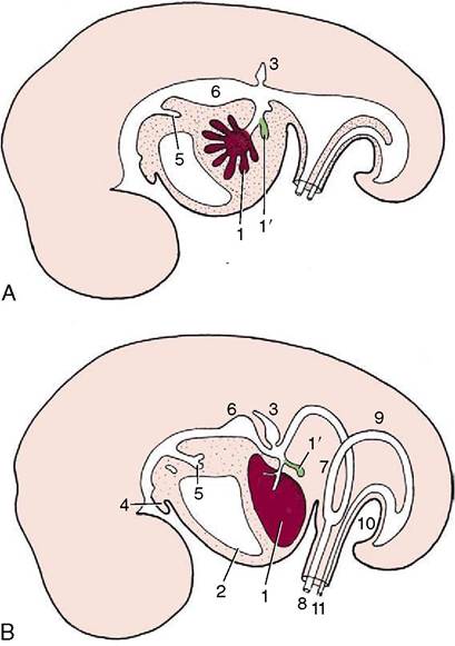
Figure 3-62 Development of the liver. A, Early development: a cranial branch (1) of the endodermal diverticulum invades the septum transversum; a caudal branch (1') forms the gallbladder and cystic duct. B, A later stage, in which the developing liver expands caudally into the abdominal cavity. 1, liver; 1, gallbladder; 2, pericardium and heart; 3, dorsal primordium of pancreas; 4, tongue; 5, tracheobronchial diverticulum; 6, stomach; 7, loop of midgut; 8, vitelline duct; 9, hindgut; 10, cloacal membrane; 11, allantoic stalk.
arrive here from the extraembryonic membranes. Very soon a three-dimensional spongework of hepatic cell-cords and plates is formed, surrounded on all sides by thin-walled blood vessels, which is a precocious realization of the adult arrangement. Attenuation of the connection between the liver and the gut forms the lesser omentum.
The growth of the liver, extremely rapid in younger embryos, is a major factor in the temporary herniation of the midgut (see further on). Although its growth slows later, the liver remains disproportionately large (by comparison with that of the adult) until well after birth. One relevant factor is the exercise of an erythropoietic activity before birth that is later relinquished. The secretory and metabolic functions are established by midterm in the human fetus.
The pancreas arises from the same portion of the foregut as the liver. There are initially two primordia: one is dorsal and the second is ventral and associated with the hepatic outgrowth (Figure 3-63). These later fuse, allowing combination of the two duct systems, following which one or the other may lose its connection with the gut. The islet tissue develops by budding from the ducts. Both endocrine and exocrine components are competent well before birth.
The celiac artery is associated with the postpharyngeal part of the foregut.