THE PHARYNX
Many details of the development of the pharyngeal region are more appropriately considered in Chapters 2 and 6. The pharynx is initially dorsoventrally flattened and widest immediately behind the oral plate, but the initial form is altered by the unequal growth of the mesoderm flanking the endodermal tube (Figure 3-60).
This mesoderm forms serial thickenings, the pharyngeal (branchial) arches, which protrude into the pharyngeal lumen and bulge on the surface of the neck. The internal modeling of the lumen defines a series of pouches with which corresponding grooves coincide externally (see Figure 3-60). The number of arches (and therefore of pouches) is disputed. It is most commonly assumed that five arches exist, representing the first four and the sixth of the somewhat longer series found in other vertebrates. Each arch develops an internal skeleton and musculature with which a particular cranial nerve is associated; the fates of these are tabulated elsewhere (p. 57). Each pouch has a specific fate (see Figure 6-5). The features of immediate interest include the contributions of the first and, possibly, the second pouches to the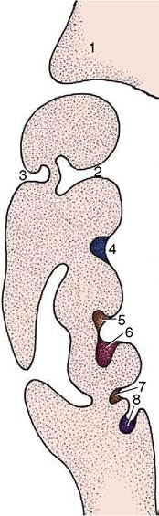
Figure 3-60 Dorsal section of the left side of the pharynx showing the development of the pharyngeal arches and pouches. 1, Maxillary process; 2, pharyngotympanic tube (future auditory tube); 3, external auditory meatus; 4, palatine tonsil (in tonsillar sinus); 5, parathyroid gland III; 6, thymus; 7, parathyroid gland IV; 8, ultimobranchial body.
cavity of the middle ear, which is a fate revealed in the adult by the site of entry of the auditory tube into the nasopharynx. The ventral part of the second pouch forms the tonsillar sinus, a landmark providing some clue to the former position of the oral plate.
The outgrowth of the lower respiratory tract at the caudal limit of the pharynx is considered in the following chapter.