THE CAVITY OF THE LARYNX
The cavity of the larynx may be divided into three sections arranged in series (Figures 4-13 and 18-35). The vestibule extends from the laryngeal entrance to the rostral margin of the arytenoid cartilages and vocal folds.
The glottic cleft is bounded by the arytenoid cartilages dorsally and the vocal folds ventrolaterally and can be varied in size. The third, infraglottic, cavity is of fixed dimensions and leads smoothly to the lumen of the trachea (Figure 4-14).The structures bounding the entrance to the larynx (aditus laryngis) project into the lumen of the pharynx; they may protrude through the intrapharyngeal ostium into the nasopharynx, where they may be grasped by the free margin of the soft palate and its continuation by the palatopharyngeal arches. The rostral part of the wall of the entrance is provided by the epiglottis, the lateral parts by the (aryepiglottic) folds extending between the epiglottis and the arytenoid cartilages, and the caudal part by the corniculate processes of the ary-
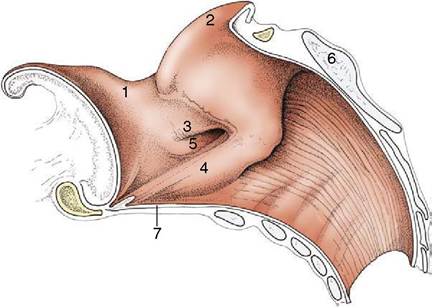
Figure 4-13 Median section of the equine larynx. 1, Epiglottis; 2, corniculate process of arytenoid cartilage; 3, vestibular fold; 4, vocal fold; 5, laryngeal ventricle; 6, lamina of cricoid cartilage; 7, cricothyroid ligament.
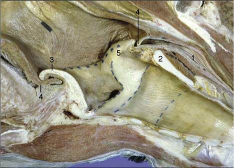
Figure 4-14 Sagittal section of junction of pharynx with larynx (horse). 1, Esophagus; 2, cricoid lamina; 3, epiglottis; 4, palatopharyngeal arch; 5, corniculate process of arytenoid cartilage.
tenoid cartilages. The interior of the vestibule may present a number of important features, but none of these is found in every species. In some animals a vestibular fold runs roughly parallel to the vocal fold but at a more rostral level (Figure 4-13/5).
This fold pairs with an outpouching of the mucosa to form a ventricle or diverticulum that is entered between the vestibular and vocal folds (Figure 18-35). These features are especially prominent in the horse and receive more attention later. The mucous membrane bounding the vestibule is tightly adherent to the epiglottic and arytenoid cartilages but is looser elsewhere where it rests on fat.The glottic cleft (rima glottidis) is narrower than the vestibule: the dorsal part is bounded by the vocal processes and adjacent parts of the arytenoid cartilages, and the ventral part is bounded by the vocal folds (the folds and the arytenoid cartilages constitute the glottis). The cleft, laterally compressed and diamond-shaped, varies in dimensions and disappears when the glottis is closed. The vocal folds run caudodorsally from the rostral part of the laryngeal floor to their attachments on the arytenoid cartilages. Each fold contains a ligament in its free margin and, lateral to this, the vocalis muscle, which is surrounded on most sides by fat. The vestibular folds, when present, have a similar construction but form no part of the glottis in the strict sense. The mucosa is tightly adherent to the arytenoid cartilages and along the free margin of the folds; it is much looser elsewhere.
The infraglottic cavity has few features of interest: its form reflects that of the cricoid cartilage. It may be slightly reduced in size where it continues into the trachea. The mucosa is relatively firmly attached.
The laryngeal mucous membrane contains numerous mucous glands (especially massed within the ventricles when these are present) and also lymphoid aggregations (especially in the infraglottic region). It is surfaced by an epithelium whose character varies from region to region according to its use. This epithelium is stratified squamous about the entrance, where it risks abrasion from the passage of food, and also on the free edges of the folds, which at times are abruptly brought together; elsewhere it is pseudostratified and ciliated like the epithelium lining most respiratory passages. The sensory innervation is from the cranial and caudal (recurrent) laryngeal nerves; the boundary between the territories coincides with the glottis.