THE MUSCULATURE
In addition to the extrinsic laryngeal muscles that pass between this organ and the pharynx, tongue, hyoid
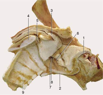
Figure 4-11 Median section of the equine larynx after removal of the mucosa.
1, Epiglottic cartilage; 2, sectioned body of thyroid cartilage; 3, corniculate process of arytenoid cartilage; 4, sectioned lamina of cricoid cartilage; 5, cricoarytenoid joint; 6, ventricularis; 7, vocalis; 8, laryngeal ventricle; 9, tracheal rings.
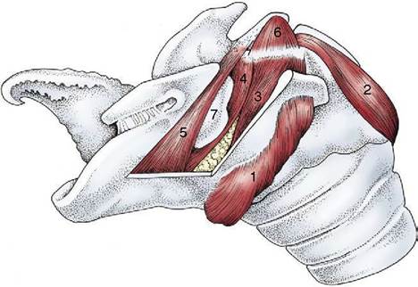
Figure 4-12 Intrinsic muscles of the equine larynx. 1, Cricothyroideus; 2, cricoarytenoideus dorsalis; 3, cricoarytenoideus lateralis; 4, vocalis; 5, ventricularis(4z5/ thyroarytenoideus); 6, arytenoideus transversus; 7, laryngeal ventricle.
bone, and sternum, a suite of small, paired, intrinsic muscles connects the laryngeal cartilages and influences their mutual relations (Figure 4-12).
One of these muscles, the cricothyroideus (Figure 4-12/7), is somewhat set apart from the rest through its superficial position and its innervation by the cranial laryngeal nerve, a branch of the vagus. It runs between the lateral surfaces of the thyroid lamina and cricoid arch ventral to the cricothyroid joint; on contraction it approximates these attachments and thus carries the dorsal part of the cricoid (and the attached arytenoid cartilages) caudally, which tenses the vocal folds.
The other muscles lie more deeply, attach to the arytenoid cartilage, and are innervated by the caudal (recurrent) laryngeal branch of the vagus nerve. The Cricoarytenoideus dorsalis (Figure 4-12/2) arises from the dorsal surface of the cricoid lamina, and its fibers converge rostrolaterally to insert on the muscular process of the arytenoid cartilage.
On contraction it abducts the vocal process and thereby the vocal fold and so widens the glottis. The cricoarytenoideus lateralis (Figure 4-12/5) takes origin from the rostroventral part of the cricoid arch and passes dorsally to an insertion on the muscular process. It is therefore an adductor of the vocal processes and thus narrows the glottis. The thyroarytenoideus arises from the cranial part of the laryngeal floor (chiefly the thyroid cartilage) and runs dorsocaudally to insert on the muscular process and adjacent part of the arytenoid cartilage. In certain species (horse and dog included) it is divided into two units, a rostral ventricularis (Figure 4-12/5) and a caudal vocalis (Figure 4-12/7), which occupy the vestibular and vocal folds. This muscle adjusts the tension of the fold(s) and forms part of the sphincter arrangement. The arytenoideus transversus (Figure 4-12/d) runs from the muscular process of the arytenoid cartilage to a median raphe (sometimes containing the interarytenoid nodule); some fibers may cross the midline to reach the arytenoid cartilage of the other side. It approximates the arytenoid cartilages and completes the sphincter.