The Corticospinal Tract Has a Massive Lateral Component Controlling the Distal Musculature and a Minor Medial Component Controlling the Axial and Proximal Musculature
The corticospinal tract axons primarily originate from cells located in the motor cortices of the frontal lobe of the cerebral hemisphere (Figure 10-4). All cells contributing to the tract are located in the fifth of the six histological layers of cortical tissue.
Along their route from the cerebral cortex, these corticospinal axons pass through the internal capsule of the forebrain, the cerebral peduncles on the ventral surface of the midbrain, and the pontine nuclei within the ventral pons, and they emerge on the ventral surface of the medulla, adjacent to the midline, as the pyramids. These appear pyramid-shaped in cross section, partly inspiring the name pyramidal tract for axons that pass through them.As the corticospinal tract axons reach the Spinomedullary border, the vast majority (85%-90% in primates) cross the mid- Iine at a structure called the pyramidal decussation (Figure 10-4). The crossing axons then form the lateral corticospinal tract, located in the lateral spinal cord white matter, and synapse within lateral regions of the spinal cord gray matter (Figure 10-5). As noted earlier, the lateral regions of the spinal gray matter contain premotor and α motor neurons that primarily control the distal flexor musculature of the extremities that parti cipate in skilled, manipulative, usually voluntary movements. Given this organization, damage to the motor cortices on one side of the body has devastating effects on voluntary skilled movement of the distal flexor musculature on the opposite side of the body. A much smaller percentage of axons traveling in the medullary pyramid do not cross the midline at the pyramidal decussation and remain on the same side of the body to form the much smaller ventral corticospinal tract (see Figure 10-4). The axons of this tract are located in more medial regions of the spinal white matter and synapse in more medial regions of the spinal gray matter that control the axial and proximal postural musculature (see Figure 10-5).
'Γhe ventral corticospinal tract provides a direct means of volun-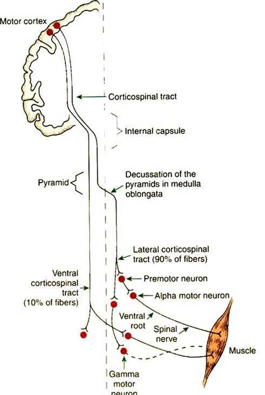
FIGURE 10-4 Corticospinal tract is a direct route primarily from the motor cortices to the contralateral spinal cord gray matter. Most axons of the tract synapse on premotor neurons of the intermediate zone, but some, depending on species phylogeny, synapse directly on α and γ lower motor neurons. About 90% of the axons of the tract cross the midline at the Spinomedullary border to form the lateral corticospinal tract, and about 10% remain on the same side to form the ventral corticospinal tract.
tary control over muscles (hat are normally involved in a subconscious antigravity function.
The ability of the corticospinal tract to control the most dexterous, skilled movements of the body derives from the synaptic termination pattern of several of its axons. The greater the number of synapses between a neuron in the motor cortices and an c/. motor neuron in the spinal cord ventral horn, the greater is the number of α motor neurons activated and the less precise the control of the musculature. This is true because each neuron that is excited in the pathway usually activates several postsynaptic neurons. Corticospinal axons bypass synapsing with neurons of the brainstem motor pathways to the cord, but more significantly, some corticospinal axons can bypass synapsing with premotor neurons of the
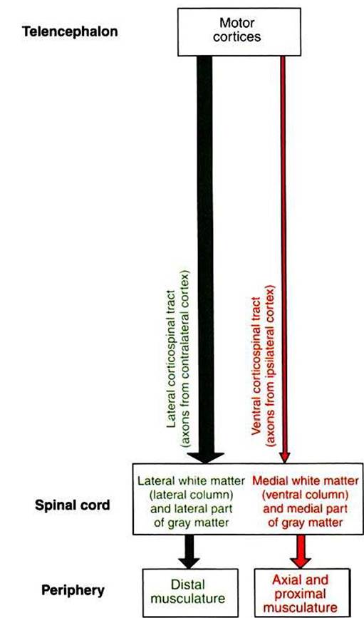
FIGURE 10-5 ■ Somatotopic relationship of corticospinal tract components. As with the descending brainstem motor pathways, the corticospinal tract can be divided into components that respectively travel in more lateral or more medial regions of the spinal cord white matter. The massive and laterally located lateral corticospinal tract synapses in more lateral regions of the spinal cord gray matter that control the distal limb musculature.
The axons of this tract originate from the contralateral motor cortices.The much smaller ventral corticospinal tract, whose axons originate from the ipsilateral motor cortices, travels in more medial regions of the spinal white matter and synapses in more medial regions of the spinal gray matter that control the axial and proximal musculature.spinal gray matter, contacting a motor neurons directly. Therefore a given corticospinal neuron can ultimately control smaller numbers of α motor neurons and a smaller complement of the musculature. This permits increased fractionation of movement, the increased independence of the actions of different muscles (e.g., the ability to move individual lingers instead of all the fingers together). The proportion of corticospinal neurons making monosynaptic connections with spinal α motor neurons is related to phylogeny. There are no such connections in cats, a small proportion in monkeys, a larger proportion in the anthropoid apes, and a still larger proportion in humans, where the most skilled, fractionated, manipulative movements take place.
As noted, cranial nerve nuclei in the brainstem contain lower motor neurons controlling muscles of the face and head. A significant complement of the axons that leave the motor cortices to form the corticospinal tract will not continue to the spinal cord, but will leave the tract to synapse at the cranial nerve motor nuclei of the brainstem. This complement of axons is referred to as the Corticobulbar tract (“bulb” being an archaic term for portions of the brainstem). A given cranial nerve nucleus generally receives significant cortico- bulbar input from both cerebral hemispheres.
As discussed earlier, the motor cortices send axons to synapse in the brainstem reticular nuclei and in the red nucleus of the mesencephalon as part of an indirect route (e.g., cortico- reticulospinal, cortico-rubrospinal) for cortical control of spinal lower motor neurons. However, such cortical projections to brainstem nuclei, other than to cranial nerve motor nuclei, are not usually viewed as belonging to the Corticobulbar tract. Another significant example ol such a projection synapses on diffuse aggregates of cells in the ventral pons called the pontine nuclei. The cells of the pontine nuclei then project to the contralateral cerebellum. It is thought that this cortical route to the cerebellum is used to inform the cerebellum of movement intended by the cerebral cortex, so that if the actual movement is not the one intended, the cerebellum can initiate appropriate adjustments.