The Craniolateral Muscles
This group comprises the tibialis cranialis, peroneus tertius, and long and lateral digital extensors. All are flexors of the hock, and those that proceed farther are extensors of the digit.
The tibialis cranialis arises from the lateral condyle and tuberosity of the tibia and continues distally, closely applied to the bone (Fig. 24.8/3). The insertion tendon begins just above the level of the hock and passes through a split in the tendon of the peroneus tertius before dividing itself. The larger dorsal branch continues to the metatarsal tuberosity. The smaller medial branch diverges to cross the medial collateral ligament before inserting on the combined first and second tarsal bones (Fig. 24.11). When the muscle contracts, it presses on the seat of spavin. Although the tibialis cranialis appears to be a flexor of the hock, it is difficult to be certain of its function. According to one view, its prime role is to counteract the bending moment applied to the tibia by the action of other muscles and by gravity.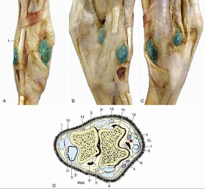
FIG. 24.11 (A to C) Illustrations of hock. (A) Dorsal view of right hock. 1, Long digital extensor; 2 and 3, Iaterodorsal and mediodorsal pouches of tarsocrural joint, respectively, filled with latex. (B) Lateral view of right hock. 1 and 2, Lateral plantar and laterodorsal pouches of tarsocrural joint, respectively, filled with latex. (C) Medial view of right hock. 1 and 2, Mediodorsal and medioplantar pouches of tarsocrural joint, respectively. (D) Bursae, tendon sheaths, and joint pouches of the left hock. 1, Superficial digital flexor; 2, calcaneus; 3, lateral deep digital flexor and tibialis caudalis (combined tendon in B); 3', tendon of medial deep digital flexor; 3", tarsal sheath; 4, cranial branch of medial saphenous vein; 5, long digital extensor;
6, peroneus tertius; 7, tibialis cranialis and underlying bursa; 8 and 8', dorsal and medioplantar pouches of tarsocrural joint, respectively; 9 and 9', medial and lateral collateral ligaments (superficial parts), respectively; 10, long plantar ligament; 11, plantar nerves and saphenous vessels; 12, cranial tibial vessels and deep peroneal nerve; 13, lateral digital extensor; 14, caudal cutaneous sural nerve and lateral saphenous vein; 15, talus; Cr., cranial; Med., medial.
The peroneus tertius is almost exclusively tendinous (Fig. 24.8/4). It arises from the lower end of the femur together with the long extensor, and for much of its course it is recessed in the deep surface of that muscle. It bifurcates at the hock with the lateral branch inserting on the calcaneus and fourth tarsal bone, and the dorsal one on the proximal part of the third tarsal and third metatarsal bones (Fig. 24.13/1). The tendon links the actions of the stifle and hock joints. Rupturing of this muscle (see Fig. 24.16A) enables extension of the hock while retaining a flexed stifle, which is a combination of movements normally impossible.
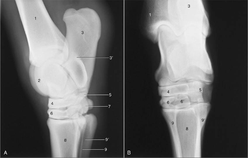
FIG. 24.12 (A) Lateral and (B) dorsoplantar radiographs of the hock joint. 1, Tibia; 2, talus; 3, calcaneus; 3', sustentaculum tali; 4, central tarsal; 5, fourth tarsal; 6, third tarsal (in B, superimposed on tarsal 1 and 2); 6', plantar projection of third tarsal; 7, tarsals 1 and 2; 8, large metatarsal bone; 9 and 9', medial and lateral splint bones, respectively.
The long digital extensor, the largest muscle of the group, arises in common with the peroneus tertius by a short tendon. This is soon succeeded by a broad belly that covers the tibialis cranialis (Fig. 24.14/5). The insertion tendon begins in the lower leg and continues to the extensor process of the distal phalanx, with passing attachments to the proximal and middle phalanges. It is joined by the smaller tendon of the lateral digital extensor (Fig. 24.14/6) near the middle of the cannon. As it descends on the dorsal surface of the limb, it is surrounded by a synovial sheath from retinacula where it crosses the hock. This muscle is capable of flexion of the hock and extension of the digit.
The lateral digital extensor runs between the long extensor and the deep flexor on the lateral aspect of the limb. It arises from the lateral collateral ligament of the stifle and adjacent parts of both tibia and fibula and ends by joining the long extensor tendon. Its tendon is also held down by retinacula and protected by a synovial sheath where it crosses the hock. A very small, short digital extensor muscle (extensor digitalis brevis) occupies the angle between the converging tendons of the larger muscles (Fig. 24.14/10). It is of no importance (Table 24.4).
All muscles of the craniolateral group are supplied by the peroneal nerve.