The Caudal Muscles
This group comprises the popliteus, whose action is confined to the stifle, and the gastrocnemius, soleus, and superficial and deep digital flexors, which all extend the hock with the last two also flexing the digit.
The popliteus is a relatively small triangular muscle placed directly over the caudal aspect of the stifle joint (Fig. 24.15B/7). It arises from the lateral condyle of the femur and inserts on the caudomedial border of the tibia. The popliteus flexes the stifle and rotates the leg inward.
The gastrocnemius, the most superficial and largest muscle of the group, arises by two heads from the supracondylar tuberosities of the femur (Fig. 24.15/1). The heads, which are first covered by the hamstring muscles, soon unite in a single strong tendon that is a major component of the calcanean tendon. The gastrocnemius tendon inserts on the point of the hock where it is covered by the tendon of the superficial flexor. To attain this deep position, it must first wind around the lateral border of the flexor tendon, where it is cushioned by the interposition of a synovial bursa (see later). Theoretically, the gastrocnemius is a flexor of the stifle and extensor of the hock, but because the tendons of the peroneus tertius and superficial flexor ensure that these joints extend or flex together, it is difficult to envisage its action. It has been asserted that its prime function is comparable to that of the tibialis cranialis—that is, adjustment of the load on the tibia. A ribbon-like soleus runs from the head of the fibula to the gastrocnemius tendon but is of no importance.
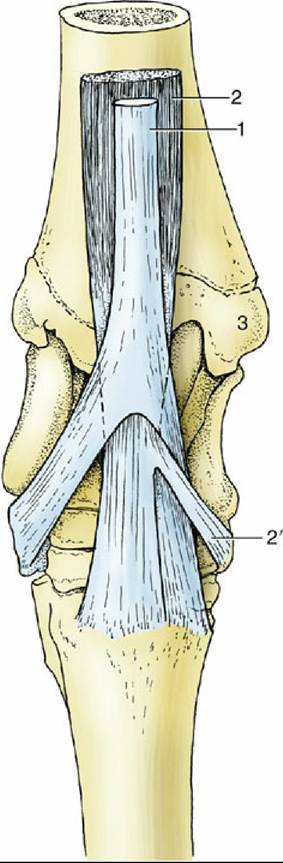
FIG. 24.13 The insertion of the flexors of the right hock, dorsal view. 1, Peroneus tertius, splitting into dorsal and lateral branches; 2, tibialis cranialis, splitting into dorsal and medial (cunean, 2) branches; 3, medial malleolus.
The superficial digital flexor (Fig. 24.15B/3) is largely tendinous, although it has a slightly greater content of flesh than the peroneus tertius. The highly diminished muscle fibers dampen the vibrations in the tendinous part to prevent overheating and tissue damage. It arises from the supracondylar fossa of the femur under cover of the gastrocnemius and, twisting around the medial surface of the tendon of that muscle, passes toward the calcanean tuber, where it expands to form a cap. The medial and lateral edges attach at the tuber, and the main part continues over the plantar aspect of the hock to enter the cannon, followed by insertion on the first and second phalanges in similar fashion to the superficial flexor of the forelimb. A considerable synovial bursa protects the expanded tendon where it caps the tuber, and also extends proximally between the flexor and gastrocnemius tendons, where they wind around each other (Fig. 24/10/1'). A second, smaller, subcutaneous bursa may form over the expanded tendon where it caps the calcaneus ("capped hock"). Both bursae usually communicate and are liable to inflammation and distention. The proximal part of the muscle is a main constituent of the so-called reciprocal mechanism (p. 626). The distal part supports the fetlock and pastern joints in similar fashion to the superficial flexor of the forelimb (Table 24.5).
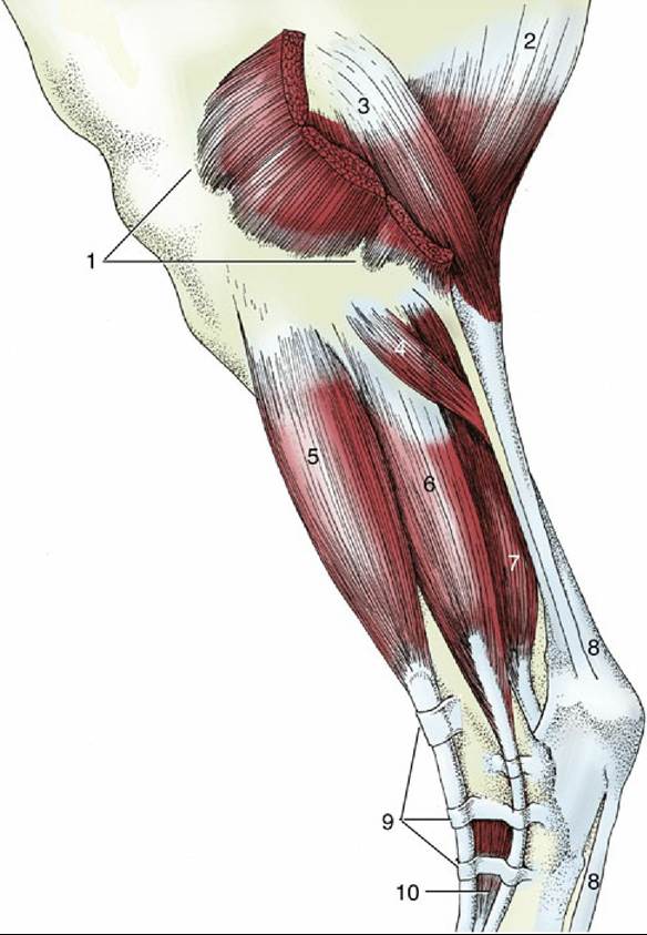
FIG. 24.14 The stifle and leg, lateral view. 1, Distal divisions of biceps; 2, semitendinosus; 3, gastrocnemius; 4, soleus; 5, long digital extensor; 6, lateral digital extensor; 7, deep digital flexors; 8, superficial digital flexor; 9, proximal, middle, and distal extensor retinacula; 10, extensor digitalis brevis.
The deep digital flexor arises by three separate and individually named heads—lateral digital flexor, medial digital flexor, and tibialis caudalis—which later unite to form a single stout tendon of insertion.
The medial flexor arises from the lateral condyle of the tibia but soon swings to the medial side of the leg (Fig. 24.15/5). The narrow tendon passes the hock, resting within a groove on the medial malleolus and medial collateral ligament, where it is protected by a synovial sheath. Once past the hock, the tendon unites with the tendon common to the other two bellies.» TABLE 24-4
The Cariolateral Muscles of the Leg
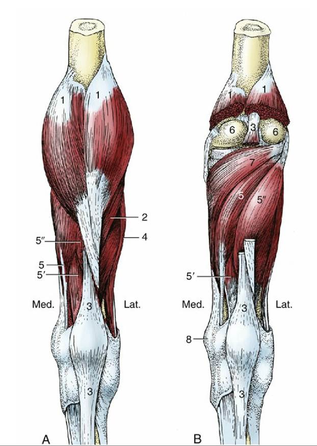
FIG. 24.15 (A) Superficial and (B) deep muscles of the right leg, caudal view. 1, Gastrocnemius; 2, soleus; 3, superficial digital flexor; 4, lateral digital extensor; 5, 5', and 5", medial and lateral deep digital flexors and tibialis caudalis, respectively; 6, femoral condyles; 7, popliteus; 8, medial malleolus; lat., lateral; Med., medial.
The lateral flexor and the tibialis caudalis have extensive origins from the caudal surface of the tibia, distal to the attachment of the popliteus (Fig. 24.15/5' and 5"). They are difficult to separate, and there is little merit in attempting the distinction because the tendons combine in the lower part of the leg. The common tendon crosses the plantar aspect of the hock over the sustentaculum tali of the calcaneus. A synovial (tarsal) sheath invests the tendon from the distal part of the leg to its junction with the tendon of the medial flexor in the upper part of the cannon (Fig. 24.11D/3 "). A further tendinous slip (the accessory ligament) that passes from the joint capsule to join the common tendon is analogous to the forelimb formation but is usually less developed and may even be absent. The distal part of the tendon comports itself similarly to the corresponding part of the deep digital flexor of the forelimb.
The deep plantar metatarsal fascia resembles the corresponding forelimb fascia and offers the same obstruction to palpation of the flexor tendons in the proximal half, and more, of the cannon.
The tibial nerve supplies all muscles of the caudal group.
The remaining structures of the metatarsus and digit closely resemble the corresponding parts of the forelimb. Certain quantitative differences have been mentioned (p. 574 and 588 and see Fig. 23.36).