» The Croup, HIP, and Thigh (see also pp. 83, 85-87, and 89.)
The habitual stance varies among breeds. The major differences are well illustrated by the German Shepherd, which tends to crouch with the back and croup sloping down toward the tail (and the hip, stifle, and hock joints markedly flexed), and the Boxer, which favors a stiffer, more upright posture (with the major joints, particularly the hock, significantly straighter).
The more upright limb appears to predispose to several common stifle disorders. In the Greyhound and other lean, short- coated dogs, the gluteal muscles such as the superficial gluteal create the croup contour. However, such details are more often obscured by subcutaneous fat or a thick coat. The major skeletal landmarks are always palpable and reveal the small angle the ilium makes with the vertebral column.The dorsal and ventral spines of the ilium are very prominent and easily palpated. The convex (iliac) crest joining these points can also be followed in its length and provides a convenient site for bone marrow biopsy in larger breeds, but is too thin to serve this purpose in smaller animals. A narrow strip of the pelvic floor bordering the ischial arch can usually be palpated between the salient tubers. In the dog the cordlike sacrotuberous ligaments, which are lacking in cats, can also be palpated. The greater trochanter of the femur is found cranial to the ischial tuber, and because its summit is very nearly level with the femoral head, it provides a good guide to the position of the joint, which is not itself palpable.
The luxation of the femur (coxofemoral) from the acetabulum is due to trauma. The symmetry of the ilium, ischial tuber, and femur may reveal luxation of the femur. This is a relatively frequent mishap with the femoral head most often displaced dorsocranially (which widens the ischiofemoral gap), but it may pass dorsocaudally or, though rarely, ventrocaudally when it engages within the obturator foramen.
The ligament of the head of the femur is especially likely to be ruptured or avulsed in full luxation but may survive subluxations. Luxation may be confirmed by rotating the thigh outward while the thumb is pressed between the trochanter and the tuber; the movement normally forces the thumb from the recess, but a luxated femur is unable to exert the necessary leverage.The hip joint possesses greater range and versatility of movement in the dog and cat than in other domestic species. The enhanced potential for abduction allows dogs to cock their legs when urinating, while the general versatility combined with the suppleness of the trunk enables both species to reach most parts of the head, neck, and thorax with the hindpaw. The articular surfaces reflect these abilities. The femoral head is an almost perfect hemisphere, marred only by the small central fovea where the intracapsular ligament (of the femoral head) inserts. It is deeply seated within the acetabular cup, which is only slightly extended by a labrum about its rim (see Fig. 2.58). There are no peripheral ligaments to limit movement, although some capsule reinforcements can be identified. The intracapsular ligament may be hypertrophied in preexisting dysplasia of the joint. In normal hips, the ligament checks the movements that endanger stability of the hip joint. The joint capsule also maintains the femoral head within the socket and prevents overextension and flexion. The fit of the femoral head within the acetabulum can be estimated from a ventrodorsal radiograph of the pelvis by measuring the Norberg angle, that is, the angle between the line connecting the centers of the femoral heads and that connecting the center of a femoral head with the cranial part of the related acetabular rim. An angle of less than 105 degrees indicates displacement and suggests dysplasia.
The blood supply to the joint capsule, the femoral neck, and the proximal epiphysis arises from an extracapsular ring formed by the lateral and medial circumflex femoral arteries, and the caudal gluteal artery.
The branches from the ring ascend the femoral neck and provide the epiphyseal arteries of the femoral head. Arteries demonstrable in the ligament of the femoral head are thought to be of little significance in the dog but make a major contribution to the supply of the femoral head of the kitten. Trauma to the femoral neck often leads to its resorption because of the limited blood supply.The most convenient access to the joint, for puncture and in surgery, is from the Craniolateral direction. An approach between the tensor and biceps muscles exposes the proximal part of the vastus lateralis (whose origin runs from just below the greater trochanter) and the gluteal muscles that clothe the joint directly. The procedure may create a minor danger of damage to the sciatic nerve and the caudal gluteal vessels.
The radiologic anatomy is very relevant to the diagnosis of the two conditions that commonly affect the joint: luxation and dysplasia. For the standard ventrodorsal radiograph (Fig. 17.1A), the supine animal must be placed with its hindlimbs drawn uniformly backward to ensure symmetrical depiction of bilateral structures. Although most features of the pelvis are too obvious to require comment, attention may be drawn to the slight lateral bowing of the canine ilia (in contrast to their parallel course in the cat). The relationship between the rim of the acetabulum and the femoral head on which it is superimposed is of the greatest importance in determining the integrity of the joint (Fig. 17.1/3). Attention is also directed to the relative radiolucency of the region (corresponding to the trochanteric fossa) between the greater and lesser trochanters of the femur, because it is sometimes misinterpreted. The less useful lateral view reveals the position of the hip joints below the first two caudal vertebrae (Fig. 17.1D).
A special position, in which the hindlimbs of the supine animal are rotated inward until the femoral trochleae and patellae face directly upward, is used for the better depiction of the contours of the femoral head when hip dysplasia is suspected.
In this view it is easier to gauge the congruence of the femoral head with the acetabulum and to recognize any flattening or distortion of its contours. Progressive deformation of the head and worsening of fit characterize the progress of the condition.The etiology of hip dysplasia, which is very common in certain larger breeds, is uncertain, but hereditary factors may be the primary drivers. It is believed that the dysplasia, which inevitably leads to osteoarthritic changes, is a consequence of the instability permitted by abnormally lax soft articular tissues. The synovitis also may lead to the accumulation of fluid in the joint and reduce the stability of the joint associated with the suction of the thin layer of the synovium between the surfaces of the head of the femur and the acetabulum.
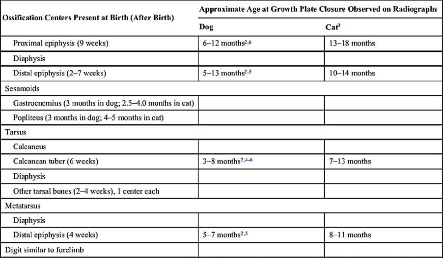
The maturation of the skeleton can be followed in radiographs obtained from young animals. In puppies there are primary ossification centers for the bodies of the ilium, ischium, and pubis and for the acetabular bone and secondary centers for the iliac crest, ischial tuber, and the border of the ischial arch. The acetabular bone is the first to lose its independence, but this is followed by the merger of the other primary centers at a comparatively early age (4—6 months). The secondary centers remain distinct until much later (15 months to 5 years for the iliac crest and 8-14 months for the ischial tuber). Fusion at the proximal extremity of the femur is completed between the 6th and 12th month (Table 17.1; see Fig. 5.74).
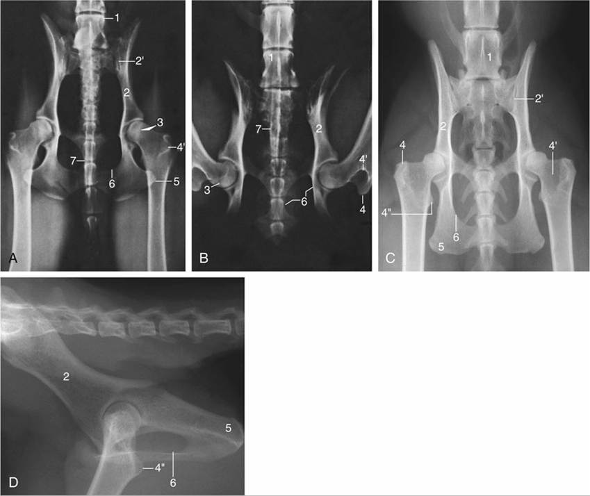
FIG. 17.1 (A) and (B) Ventrodorsal radiographic views of the canine pelvis with (A) extended and (B) flexed hip joints. (C) and (D) Radiographs of the feline pelvis in (C) ventrodorsal and (D) lateral views; D is taken of a specimen. 1, Last lumbar vertebra (L7); 2, shaft of ilium; 2', sacroiliac joint; 3, dorsal border of acetabulum superimposed on the femoral head; 4, greater trochanter; 4', trochanteric fossa; 4", lesser trochanter; 5, ischial tuber; 6, obturator foramen; 7, os penis superimposed on vertebrae.
The shaft of the femur is so deeply embedded among the muscles of the thigh that only a general impression of its presence may be obtained on palpation (Fig. 17.2/9). Despite this protection, the femur is the most commonly fractured bone, and most breaks occur about or below midshaft level. Such fractures are often complicated by considerable overriding because the lower fragment is commonly displaced caudally by the pull of the gastrocnemius. They are often repaired by intramedullary pinning, a procedure usually requiring direct exposure of the break, which is easily achieved through a lateral approach. The incision of the fascia lata followed by reflection of the biceps, whose cranial margin is often palpable through the skin, exposes the vastus lateralis and opens a path to the bone (Fig. 17.2/8-10).
Although the caudal thigh muscles appear to lend themselves to intramuscular injection, they should be avoided for this purpose because of possible damage to the sciatic nerve; a better alternative is injection into the muscles of the back.
The gluteal muscles have been described (Table 17.2). Caudal to these, the cat presents the gluteofemoral, a long and relatively strong muscle that arises from the second to fourth caudal vertebrae and runs caudal to the superficial gluteal muscle and cranial to the biceps to insert lateral to the patella in the fascia lata. It retracts the hindlimb and may also draw the tail to the side. The biceps femoris covers the abductor cruris caudalis, a small, thin muscle strap that emerges over the lateral head of the gastrocnemius in the lower leg.
The most important palpable structure of the thigh is the femoral artery (Fig. 17.2/2), which is subcutaneous on the medial aspect of the limb toward the groin. It lies within the femoral triangle, a pyramidal space whose base lies toward the vascular lacuna (the passage to and from the abdomen for the femoral artery and vein) and whose tip is closed distally by the convergence of the sartorius and pectineus muscles that form its cranial and caudal walls.
The pectineus muscle forms so obtrusive a fusiform swelling that it immediately guides the fingers to the femoral artery, which is the first choice for taking the pulse. The femoral artery's name changes to the popliteal artery once it reaches the popliteal fossa on the medial aspect of the femur (Fig. 17.3/1 and 2). The femoral vein is less conspicuous but is easily found along the caudal border of the artery and is convenient for intravenous injection in the supine, anesthetized subject. The saphenous artery (Fig. 17.3/4) branches from the concealed part of the femoral artery but soon becomes subcutaneous and runs over the medial aspect of the thigh toward the stifle. Both it and a large, more proximal branch (running caudally toward the gracilis) may be palpated.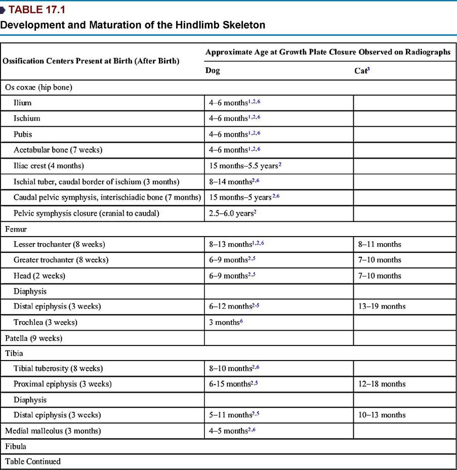
1 Based on Chapman WL: Appearance of ossification centers and epiphyseal closures as determined by radiographic techniques, J Am Vet Med Assoc 147:138-141,1965.
2 Based on Hare WCD: The age at which epiphyseal union takes place in the limb bones of the dog, Wien Tierarztl Monatsschr 9:224-245, 1972.
3 Based on Smith RN: Fusion of ossification centers in the cat, J Small Anim Pract 10:523-530, 1969.
4 Based on Smith RN and Allcock J: Epiphyseal fusion in the Greyhound, Vet Rec 72:75-79, 1960.
5 Based on Sumner-Smith G: Observations on the epiphyseal fusion of the canine appendicular skeleton, J Small Anim Pract 7:303-311, 1966.
6 Based on Ticer JW: Radiographic technique in small animal practice, Philadelphia, 1975, Saunders, p. 101. From de Lahunta A and Habel RE: Applied veterinary anatomy, Philadelphia, 1986, Saunders.
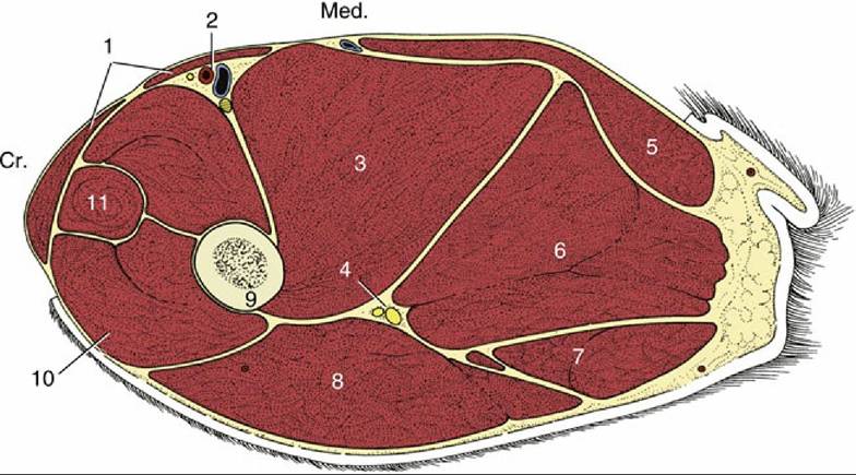
FIG. 17.2 Transverse section of the canine left thigh. Cr., Cranial; Med., medial. 1, Sartorius; 2, femoral vessels; 3, adductor; 4, sciatic nerve; 5, gracilis; 6, semimembranosus; 7, semitendinosus; 8, biceps; 9, femur; 10, vastus lateralis (of quadriceps); 11, rectus femoris.
Unlike the larger species, the dog and cat have no subiliac lymph nodes. However, the popliteal
lymph node is usually palpable within the popliteal fossa, between the distal parts of the biceps and semitendinosus as they diverge toward their insertions at the stifle (Figs. 17.4/10 and 17.5/6).
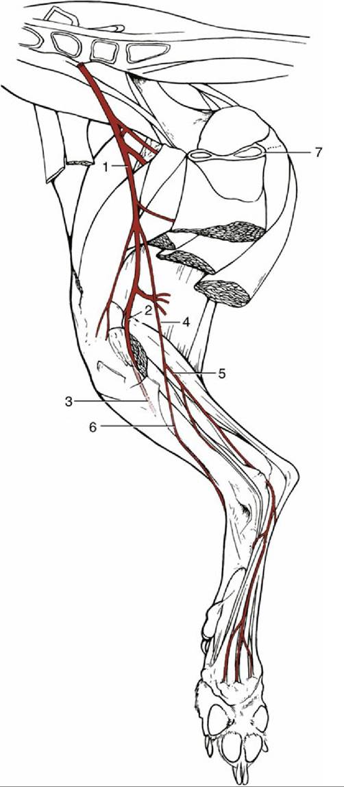
FIG. 17.3 The principal arteries of the canine right hindlimb, medial view. 1, Femoral artery (a.); 2, popliteal a.; 3, cranial tibial a. passing between tibia and fibula; 4, saphenous a.; 5 and 6, caudal and cranial branches of saphenous a.; 7, pelvic floor.