THE EPIDIDYMIS
The epididymis is a firm organ that is largely formed by the numerous convolutions of the single epididymal duct within a connective tissue matrix. It is attached along one of the longer borders—dorsal in the dog,
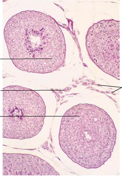
Figure 5-39 Testis (dog) (140?).
1, seminiferous tubules (showing spermatogenesis); 2, interstitial tissue with androgen-producing (Leydig) cells.
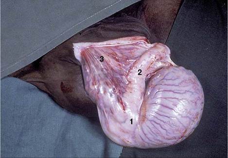
Figure 5-40 Testis (horse). 1, Head of epididymis; 2, body of epididymis; 3, pampiniform plexus.
caudomedial in the bull—of the testis and usually spreads some distance over both poles (Figure 5-40). It is conventionally divided into three parts—head, body, and tail—but these rather arbitrary divisions do not always correspond to functional distinctions.
The head (Figure 5-38Z5,) is firmly attached to the testicular capsule. It receives the efferent ductules, which immediately or after some coiling join to form the wider epididymal duct (Figure 5-38Zd'). The body may be less completely attached to the surface of the testis, and in that case an intervening space (testicular bursa, homologous with the ovarian bursa) is created (see Figure 5-41Z5). The tail is firmly attached to the testis by a ligament (proper ligament of the testis) and also to the parietal layer of the enveloping peritoneal sac by the ligament of the tail of the epididymis (Figure 5-41Z7,8). The tail finally tapers, and the duct emerges to continue as the deferent duct (see Figure 5-41Z4). The epididymis appears spongy in section because the coiled duct is inevitably cut across many times.