THE ESOPHAGUS
The esophagus at first lies between the trachea and the cervical muscles but soon deviates to the right, a posi-
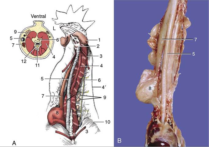
Figure 37-15 Ventral view of the dissected neck.
A, Schematic. The inset shows a transverse section through the middle of the neck. B, Detail of neck with crop. 1, Wattle; 2, larynx; 3, sternothyroideus, cut; 4, cervical muscles; 4', cervical nerve; 5, trachea; 6, jugular vein and vagus; 6', internal carotid arteries; 7, esophagus; 8, crop; 9, thymus; 10, pectoralis; 11, vertebra; 12, spinal cord.tion it maintains throughout the remainder of the neck, although both it and the trachea are quite movable (Figure 37-15, A-B). This topography makes it essential that a crop needle be introduced into the esophagus for gavage feeding or oral medication from the left side of the beak. Approach from the right side contains a high risk of perforating this thin-walled tube. At the thoracic inlet the ventral wall of the chicken’s esophagus is greatly expanded to form the crop (Figure 37-15/8), which bulges farther to the right and lies against the breast muscles. In most birds, including ducks and geese, the crop is merely a fusiform enlargement of the esophagus. Both cervical esophagus and crop are subcutaneous and palpable, ideally placed for surgery (foreign bodies, impaction) but vulnerable to laceration. The crop stores food for short periods when the muscular stomach is full. In species like owls, gulls, and penguins, which have no crop, food enters directly into the proventriculus. In piscivorous birds, fish can often be seen stretching from the proventriculus and projecting out of the beak without causing any choking or discomfort. Within the body cavity the esophagus passes over the bifurcation of the trachea, between the ventral surface of the lungs, and the base of the heart (Figure 37-16).
It merges into the proventriculus directly to the left of the median plane. Much lymphoid tissue (esophageal tonsil) is present in the caudal segment of the esophagus of the duck.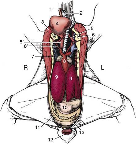
Figure 37-16 Viscera after removal of ventral body wall, ventral view. 1, Esophagus; 2, trachea; 3, pectoralis, cut; 4, crop; 5, sternotrachealis; 6, coracoid bone, cut; 7, right cranial vena cava; 8, heart; 8', common carotid artery; 8", subclavian artery; 9,9', right and left lobes of liver; 10, gizzard (its caudal blind sac); 11, duodenal loop, enclosing pancreas; 12, vent; 13, one of the ceca.
The esophagus is capable of great distention; its lamina propria contains mucous glands whose secretion lubricates the passage of the bolus. There is little chemical activity in the esophagus and crop, although salivary amylase may initiate carbohydrate digestion.
During brooding, the large symmetrical crop of both male and female pigeons elaborates a crumbly material (crop milk) consisting of desquamated lipid- laden epithelial cells; mixed with ingested food, it is regurgitated and fed to the nestlings in the first days after hatching.