» The Esophagus, Trachea, and Thymus (see also pp. 110-111, 147-148, and 250-251)
The esophagus enters the thoracic cavity to the left of the trachea but gradually assumes a median position above the trachea within the cranial mediastinum, where it is related to the left subclavian artery, which intervenes between it and the left lung (see Fig.
13.14). It continues dorsal to the trachea and subsequently to the left principal bronchus, where it crosses the heart before passing between the aorta and the azygos vein. Inclusion between these vessels and perhaps also the slight rise over the tracheal bifurcation predispose this part of the esophagus to obstruction by foreign bodies. A potentially more serious interference may be provided by the anomaly in which the right aortic arch persists as part of a constricting ring composed of the aorta to the right, the ligamentum arteriosum dorsally, and the pulmonary trunk and right pulmonary artery to the left (see Fig. 7.2D). More caudally, the esophagus rests on the left atrium and then on the accessory lobe of the right lung before reaching the hiatus in the diaphragm below the 10th thoracic vertebra. A slight narrowing here provides another site for obstruction. The chief blood supply from the bronchoesophageal artery is supplemented by direct branches from the aorta; the most caudal stretch is supplied by branches of the left gastric artery.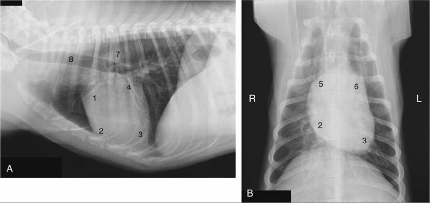
FIG. 13.17 (A) Lateral and (B) ventrodorsal views of the position of the canine heart. 1, Right auricle; 2, right ventricle; 3, left ventricle; 4, left atrium; 5, right atrium; 6, pulmonary trunk; 7, aorta; 8, trachea; L, left;
R, right.
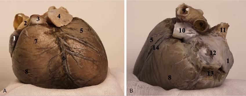
FIG. 13.18 Heart of the dog, (A) auricular surface and (B) right side. 1, Right auricle; 2, aorta; 3, pulmonary trunk; 4, left auricle; 5, left ventricle; 6, interventricular paraconal groove and paraconal interventricular branch of the left coronary artery; 7, conus arteriosus of the right ventricle; 8, right ventricle; 9, pulmonary veins; 10, caudal vena cava; 11, cranial vena cava; 12, sinus venarum cavarum.
Cranial to the heart, a surgical approach to the esophagus is easier from the left. The right approach is favored at the level of the heart because the azygos vein may be ligated with impunity, unlike the aorta to the left. The caudal section is equally approachable from either side.

FIG. 13.19 Corrosion cast of the dog's heart (caudal, atrial surface). 1, Aortic arch; 2, pulmonary trunk; 3, cranial vena cava; 4, impression for the intervenosus tubercle; 5, right atrium; 6, caudal vena cava; 7, vena cordis magna; 8, circumflex branch of the left coronary artery; 9, ramus interventricularis subsinosus artery and vena cordis media; 10, right atrium.
» TABLE 13.1
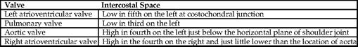
Auscultation of heart valves
The muscle is striated throughout the length of the esophagus in both dog and cat. Only the caudal section is extensively covered by serosa. Glands are present in the submucosa only in the dog. The mucosa is thrown into ridges that are predominantly longitudinal throughout the length of the esophagus of the dog but become oblique in the caudal part of the esophagus of the cat. On barium contrast radiography these oblique folds of the esophagus portray a herringbone pattern caudal to the heart in cats (Fig. 13.21B).
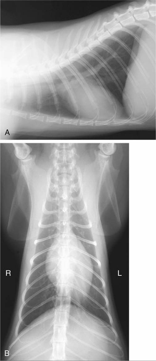
FIG. 13.20 (A) Lateral and (B) ventrodorsal radiographic views showing the position of the feline heart. The ventral ends of ribs 5, 6, and 7 lie on the heart shadow in (A).
The relationship of the trachea to the esophagus has been mentioned. The shift to a position ventral to the esophagus at the level of the aortic arch produces a caudally open angle that is a very prominent feature of lateral radiographs (Figs. 13.17/8 and 13.20). Changes in this angle may reveal abnormalities of various cranial mediastinal structures.
The relations of the trachea in this region are with the brachiocephalic trunk, common carotid arteries, and cranial vena cava. The trachea bifurcates below the fifth or sixth thoracic vertebra, where it lies above the base of the heart. It is continued by the divergent principal bronchi, of which the left is at a slightly more dorsal level despite having the esophagus resting on it.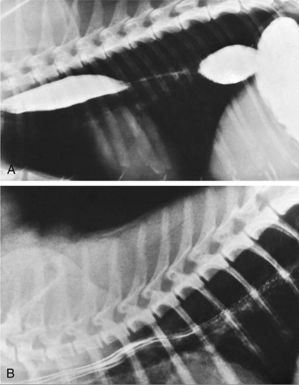
FIG. 13.21 Contrast medium in the esophagus of the (A) dog and (B) cat. Note the herringbone pattern caused by the oblique folds in the caudal part of the feline esophagus.
There are two proposed measures for the evaluation of the tracheal diameter in lateral radiographs. According to one, the tracheal diameter at the level of the third rib should be about three times the width of that rib. The other asserts that the height of the trachea should be about half that of the thoracic inlet. When the latter criterion is used, dogs with severe tracheal hypoplasia can exhibit a ratio that is only a small fraction of the value described. In this condition the deformed tracheal rings are small and thickened and have ends that meet dorsally, displacing the tracheal muscle inward, toward the lumen. It may be part of a wider "brachycephalic syndrome." Stretches of trachea reduced in size but otherwise normal have been recorded in dogs of certain large breeds. Collapse of the trachea along with abnormality of its cartilages, and sometimes also of those of the bronchi, occurs in dogs of miniature breeds.
In the dog, the thymus is confined to the thorax, where it occupies the ventral part of the cranial mediastinum, stretching from the thoracic inlet to the pericardium on which it is molded (Figs. 13.11, 13.14, and 13.22). A larger part of the thymus extends onto the left surface of the pericardium than onto the right, producing a characteristic shadow (sail sign) in dorsoventral radiographs of young dogs (those less than a year old).
The thymus consists of right and left lobes, is distinctly lobulated, is pink when fresh, and attains its greatest development at about 6 to 8 weeks. Regression begins about the fourth month but is never complete. Thymic neoplasms may compress the cranial vena cava and esophagus at the thoracic inlet.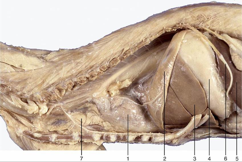
FIG. 13.22 Overview of the feline thorax, demonstrating the thymus. 1, Heart; 2, diaphragm; 3, distended stomach (with attachment of greater omentum); 4, spleen; 5, duodenum; 6, twelfth rib; 7, thymus.
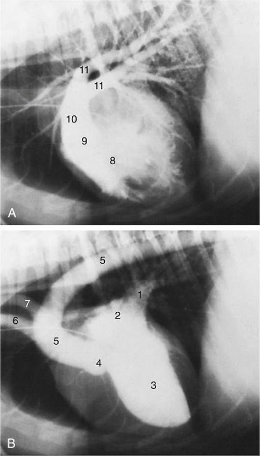
FIG. 13.23 Contrast medium in the canine (A) right and (B) left ventricles marking the great vessels. The catheter is in the cranial vena cava. 1, Pulmonary veins; 2, left atrium; 3, left ventricle; 4, position of aortic valve; 5, aorta; 6, brachiocephalic trunk; 7, left subclavian artery; 8, right ventricle; 9, position of pulmonary valve; 10, pulmonary trunk; 11, pulmonary arteries.