THE ESOPHAGUS, TRACHEA, THYMUS, AND VAGUS NERVES
The esophagus and trachea enter the thorax surrounded by a loose fascia that continues the connective tissue of the neck and provides a pathway for the spread of fluids and infection that is most relevant in connection with leaking wounds of the esophagus.
At this level the esophagus lies dorsolateral to the trachea on the left side but soon obtains a median position. Its relations include the cranial mediastinal lymph nodes and the vagus and sympathetic nerves when still close to the thoracic entrance, the aorta, thoracic duct, azygous vein, and the tracheobronchial and middle mediastinal nodes more caudally. In its final thoracic stretch, it has the important relations of the vagal trunks and the caudal mediastinal nodes (see p. 676).Postmortem, the esophagus is seen relaxed, providing no evidence of the prediaphragmatic sphincter that is sometimes alleged to exist. The part embraced by the diaphragm may be found constricted, although palpation of the hiatus in life does not support the view that the diaphragm exerts a firm grip.
The trachea, deep and compressed from side to side, first lies dorsal to the veins combining to form the cranial vena cava; it continues this relationship to its bifurcation above the right atrium, shortly after detaching the bronchus that serves the right cranial lobe. Its relations at different levels include the principal nerves within the thorax, the aorta and thoracic duct, and the tracheobronchial nodes.
The thymus has previously been encountered in the neck (p. 660 and Figure 25-24). The thoracic part fills the ventral part of the cranial mediastinum, extending at its apogee over the cranial surface of the pericardium and reaching the origin of the pulmonary trunk and the aortic arch. Involution is rarely complete, and some vestige, consisting mainly of fat and fibrous tissue, persists even in aged animals.
The sympathetic and phrenic nerves are unremarkable. The vagus nerves exhibit no special features before their division into dorsal and ventral branches that
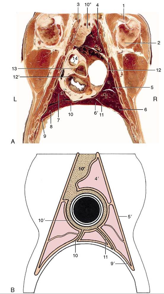
Figure 27-5 Dorsal section of the bovine thorax directly ventral to the shoulder joint. A, Actual. B, Schematized to show the asymmetry of the cranial and caudal parts of the mediastinum (stippled). 1, Biceps tendon; 2, humerus; 3, first rib; 4, cranial lobe of right lung; 4', pulmonary pleura; 5, middle lobe of right lung; 5', costal pleura; 6, 6', caudal and accessory lobes of right lung; 7, caudal part of cranial lobe of left lung; 8, caudal lobe of left lung; 9, diaphragm; 9', diaphragmatic pleura; 10,10', 10", caudal, middle, and cranial mediastinum, the last occupied by the thymus; 11, plica venae cavae; 12, 12', right and left atrioventricular valves; 13, left coronary artery arising from aortic valve; 14, pulmonary valve.
unite with their partners of the other side to form the trunks that follow the borders of the esophagus. A connection over the left face of the esophagus suggests a further rearrangement of fibers preparatory to entering the abdomen, which may be relevant to the inconsistent effects of nerve sections on gastric function. The connection sometimes suggests reinforcement of the ventral trunk at the expense of the dorsal one, and sometimes the reverse. The relationship to the caudal mediastinal lymph node(s) is of importance (see p. 676).
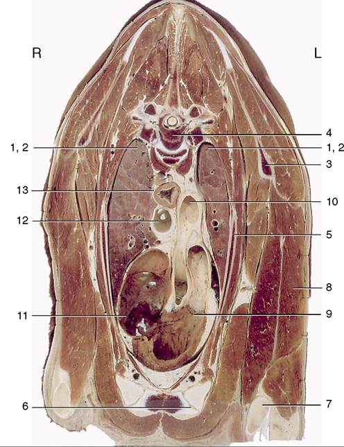
Figure 27-6 Transverse section of the bovine thorax at the level of the fourth thoracic vertebra. Note the asymmetry of the lungs. 1,2, Cranial lobes of right and left lungs; 3, scapula; 4, fourth thoracic vertebra; 5, third rib; 6, sternum; 7, olecranon; 8, long head of triceps; 9, pulmonary valve; 10, aortic arch; 11, right atrioventricular valve; 12, trachea; 13, esophagus.
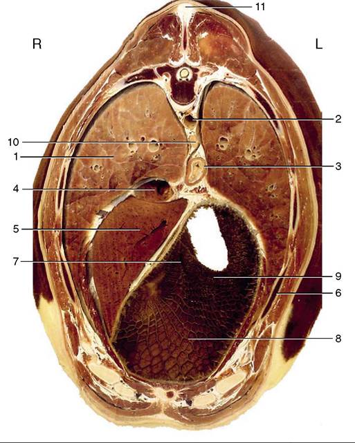
Figure 27-7 Transverse section of the bovine trunk at the level of the eighth thoracic vertebra. Note the cover to abdominal viscera provided by the ribs. 1, Caudal lobe of right lung; 2, aorta; 3, esophagus; 4, caudal vena cava; 5, liver; 6, seventh rib; 7, reticular groove; 8, reticulum; 9, ruminoreticular fold; 10, caudal mediastinal lymph node; 11, supraspinous ligament.