The Extensor Muscles
The extensor muscles constitute a large mass that fills the triangle between the scapula and humerus. The group comprises the triceps, tensor fasciae antebrachii, and anconeus and is supplied by the radial nerve.
The triceps is by far the most important extensor of the elbow. It presents three heads (Fig. 23.6B/10 and 10'). The long head arises from the caudal border of the scapula by a short aponeurosis, and the lateral and medial heads arise from the shaft of the humerus. Together they insert on the olecranon where a small bursa is inserted between the tendon and the bone. The division between the long and lateral heads is sometimes visible in thin-skinned animals. A second, acquired (adventitious) bursa is commonly found subcutaneously, over the triceps insertion and expanded part of the olecranon tuber ("capped elbow" between the long and lateral heads; Fig. 23.11/5).
» TABLE 23.2
Muscles Acting on the Shoulder Joint
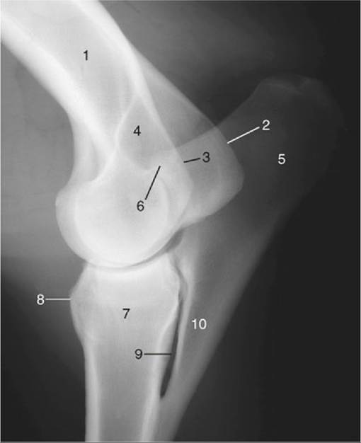
FIG. 23.9 Lateral radiograph of an elbow joint. 1, Humerus; 2, medial epicondyle; 3, lateral epicondyle; 4, olecranon fossa; 5, olecranon; 6, anconeal process of olecranon; 7, radius; 8, radial tuberosity; 9, interosseous space; 10, ulna.
The triceps is an extensor to the elbow. Because the long head spans the shoulder joint, it is theoretically available to flex this joint; however, it is probably little used for that purpose.
The tensor fasciae antebrachii (Fig. 23.8/6) is a broad, thin sheet covering the medial aspect of the triceps. Its origin is from the caudal border of the scapula and the tendon of the latissimus, while its insertion is spread between the olecranon and forearm fascia. Because it crosses both the shoulder and elbow joints, it must be considered as having a potential action at each; neither is likely to be of great importance.
The much smaller anconeus lies within the olecranon fossa, embedded within the deep face of the lateral head of the triceps and directly related to the capsule of the elbow joint. It may be supposed that its principal action is to tense the capsule, thus preventing it from being pinched between the humerus and ulna (Fig. 23.12/4 and Table 23.4).
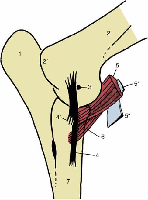
FIG. 23.10 Medial view of left elbow joint to show the eccentrically placed collateral ligament and the insertions of the biceps and brachialis. The internal tendon (5) of the biceps splits off the lacertus fibrosus (5") from the surface of the muscle. 1, Olecranon; 2, humerus; 2', medial epicondyle; 3, axis of rotation; 4 and 4', long superficial and short deep parts of medial collateral ligament, respectively; 5, biceps; 5', internal tendon of biceps; 5", lacertus fibrosus; 6, brachialis; 7, radius.