The External Ear
The external ear consists of two parts, the auricle and the external acoustic meatus (Fig. 9.24/1 and 2). The auricle, or pinna, is the "ear" as it is understood by the layperson, the part that sticks out from the head.
The external acoustic meatus is the canal that leads from the base of the auricle to the eardrum (tympanic membrane) stretched across an opening in the temporal bone.The auricle is shaped like a funnel; distally it is wide open to receive the sound, and more proximally it is rolled up to form a tube that bends medially for connection with the external acoustic meatus. The particular shape of the auricle is determined by the supporting auricular cartilage (Fig. 9.25). In most domestic mammals, the cartilage is sufficiently stiff to keep the auricle erect at all times. In many breeds of dogs and in certain other animals, the cartilage is relatively soft, allowing the auricle to collapse; even so, most dogs can prick their ears.
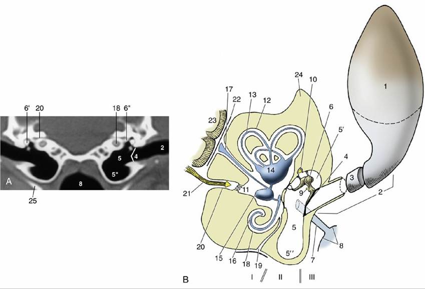
FIG. 9.24 (A) Transverse image of a 2-mm-thick computed tomography slice of the canine tympanic bullae and petrous temporal bones. (Bone settings were used.) (B) Schema of the right ear, caudal view. Note that the sizes of the structures shown are out of proportion to one another. I, Internal ear; II, middle ear; III, external ear. 1, Auricle; 2, external acoustic meatus; 3, annular cartilage; 4, tympanic membrane; 5, tympanic cavity; 5', epitympanic recess; 5", tympanic bulla; 6, auditory ossicles; 6', malleus; 6", base of stapes in vestibular window; 7, auditory tube; 8, nasopharynx; 9, chorda tympani; 10, facial nerve; 11, vestibule; 12, semicircular canals; 13, semicircular ducts; 14, utriculus; 15, sacculus; 16, cochlear duct;
17, endolymphatic duct; 18, cochlea; 19, perilymphatic duct; 20, internal acoustic meatus; 21, vestibulocochlear nerve in internal acoustic meatus; 22, meninges; 23, brain; 24, petrous temporal bone;
25, stylohyoid bone.
In domestic animals, the auricle can be turned toward the source of sound; right and left auricles can move independently so that each can focus on separate sounds.
A complex set of auricular muscles, all voluntary, is responsible for the movement of the ear. These muscles arise from various points on the skull and adjacent fasciae and attach to the base of the auricle. A flat, palpable (scutiform) cartilage rostral to the ear redirects the pull of some muscles. The auricular muscles are innervated by branches of the facial nerve.The external acoustic meatus begins where the rolled-up part of the auricular cartilage narrows and ends at the eardrum (Fig. 9.24/2). The meatus therefore has a distal cartilaginous and a more proximal osseous part. It is lined with skin that contains sebaceous and tubular ceruminous glands. The latter secrete the earwax (cerumen), which is thought to prevent dust from reaching the delicate tympanic membrane. The ear of the dog is of the most clinical interest. Unfortunately, its external acoustic meatus is curved, making passage of the straight otoscope for the examination of the proximal part of the meatus and eardrum difficult.