The Middle Ear
The middle ear is housed in the temporal bone and is essentially the small air-filled space known as the tympanic cavity (Fig. 9.24/5). It is lined with a thin mucous membrane and communicates with the nasopharynx by the auditory tube (Fig.
9.24/7). The lateral wall of the cavity incorporates the tympanic membrane (Fig. 9.24/4). The medial wall is formed by the petrous part of the temporal bone, which houses the internal ear. It contains two windows (fenestrae), closed in the natural state, through which the mechanical stimuli produced by sound waves enter the internal ear for translation into nerve impulses. The more dorsal vestibular window connects the tympanic cavity with the vestibule of the internal ear. In the live animal, the vestibular window is in contact with the stapes, the most medial of the auditory ossicles in the middle ear (Fig. 9.24/6). The other window, the cochlear window, leads to the cavity of the cochlea (Fig. 9.24/18). It is closed by the thin secondary tympanic membrane. Ventral to the two windows, the medial wall of the tympanic cavity bulges over the cochlea, forming the promontory.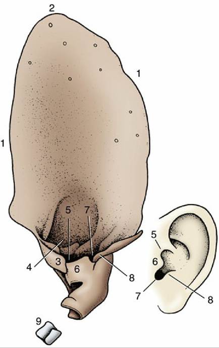
FIG. 9.25 Left auricular cartilage of dog (left) compared with human ear (right). 1, Helix; 2, apex; 3, medial crus of helix; 4, lateral crus of helix; 5, pretragic notch; 6, tragus; 7, intertragic notch; 8, antitragus; 9, annular cartilage.
The tympanic cavity may be divided into dorsal, middle, and ventral parts. The dorsal part of the tympanic cavity, the epitympanic recess, is situated dorsal to the level of the tympanic membrane and is compressed from side to side and slanted laterally. It contains the chain of three auditory ossicles and the two associated muscles. The middle part includes the tympanic membrane in its lateral wall and opens rostrally into the nasopharynx via the auditory tube.
The ventral part is an enlarged bulbous extension of the temporal bone known as the tympanic bulla (Fig. 9.24/5"). The bulla varies in prominence among species; in some it is subdivided into numerous bony cells. The function is not known with certainty but may be to improve the perception of sounds of very low and very high frequencies.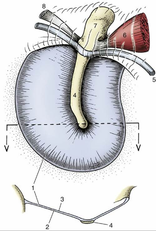
FIG. 9.26 Medial surface (top) and transverse section (bottom; location of section indicated by dotted lines and arrows) of canine tympanic membrane. 1, Tense part of tympanic membrane; 2, medial surface;
3, lateral surface; 4, handle of malleus; 5, chorda tympani; 6, tensor tympani muscle; 7, head of malleus;
8, one of the ligaments associated with the malleus.
The tympanic membrane (Fig. 9.26) is a thin partition separating the lumen of the external acoustic meatus from that of the tympanic cavity. Like the tympanic cavity, it is slanted so that its dorsal part is more lateral than its ventral part, and its surface area is thus considerably larger than that of the transected external acoustic meatus. The dog's eardrum on average measures 10 ? 15 mm; its long axis is oriented rostrocaudally. Its outer, laterally facing surface is covered with an epidermis continuous with that of the meatus, whereas its medial surface is continuous with the mucosa lining the tympanic cavity. A layer of fibrous tissue between the epidermis and mucosa firmly attaches the edges of the membrane to the osseous tympanic ring of the temporal bone. The tympanic ring is interrupted dorsally by a notch that extends onto the roof of the external acoustic meatus. The part of the tympanic membrane attached to the tympanic ring is tense; the part that closes the notch is flaccid.
The handle of the malleus (Fig. 9.26/4), the most lateral of the ear ossicles, is embedded in the medial surface of the tympanic membrane. Tension in the chain of ossicles pulls the tympanic membrane medially, hollowing its lateral surface.
The handle shines through the thin membrane and is visible as a light band (stria mallearis) when the eardrum is examined with an otoscope (see Fig.11.43 A and B).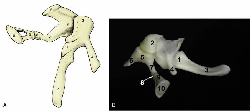
FIG. 9.27 (A) Left auditory ossicles of the horse, Craniomedial view. (B) Left auditory ossicles of the dog.
1, Malleus; 2, head of malleus; 3, handle of malleus; 4, rostral process; 5, incus; 6, short crus; 7, long crus; 8, os lenticulare; 9, head of stapes; 10, base (footplate) of stapes.
Auditory Ossicles
The transmission of sound waves across the tympanic cavity is mediated by the three auditory ossicles (Fig. 9.24/6) known, in Iateromedial sequence, as malleus, incus, and stapes (Latin names for hammer, anvil, and stirrup, from their rather fanciful resemblance to these objects).
The handle (manubrium) of the malleus (Fig. 9.27A/3 and B) is embedded in the tympanic membrane, as previously mentioned, so that the head of the malleus protrudes above the membrane by a few millimeters. The head articulates with the body of the incus, and the latter articulates with the head of the stapes by means of its long crus. The base (footplate) of the stapes sits in the vestibular window in the medial wall of the tympanic cavity.
The oscillations of the tympanic membrane transmitted by the handle of the malleus are magnified and transmitted to the vestibular window by lever action through the chain of ossicles. The base of the stapes is set in motion, causing the fluid in the internal ear to vibrate. The vibration stimulates the receptor cells in the cochlea, and sound is perceived.
The mechanism of sound transmission from the outside to the internal ear may not in fact be quite so simple. There is evidence that some sound waves are also transmitted to the fluid through the walls of the tympanic cavity and directly through the cochlear window.
The auditory ossicles are attached to the wall of the epitympanic recess by several ligaments, and their relationships can be altered by two small muscles (tensor tympani and stapedius).
These muscles tense the tympanic membrane and the chain of ossicles to decrease the amplitude of their vibrations during lower frequencies and protect the system from damage caused by sudden overload (see p. 304 for their innervation).Auditory Tube
This structure, often called the eustachian tube, connects the tympanic cavity with the nasopharynx (Fig. 9.24/8). It is short with a narrow lumen that is laterally compressed and usually collapsed. The tube is confined by an inverted cartilaginous trough except along its ventral border. The membranous wall of the horse's auditory tube evaginates through this ventral defect in the cartilaginous support to form the large, thin-walled guttural pouch dorsolateral to the nasopharynx (see p. 511).
The pharyngeal openings of the auditory tubes are located in the lateral walls of the nasopharynx and are marked by accumulations of lymphoid tissue (tubal tonsils) (see Fig. 18.11/8). The cartilage of the auditory tube extends into the medial wall of the pharyngeal opening and stiffens it. The auditory tubes allow equalization of the pressures on the two sides of the delicate eardrums. The pressure sometimes becomes unbalanced, for example, during rapid elevation changes, and its sudden restoration causes a popping sensation. The auditory tubes temporarily open each time we swallow or yawn. This opening permits the slight secretion from the goblet cells and the glands in the lining of the tympanic cavity to escape.
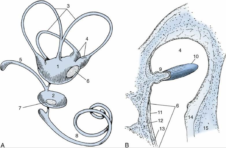
FIG. 9.28 (A) Membranous labyrinth. (B) Section of ampulla. 1, Utriculus; 2, sacculus; 3, semicircular ducts; 4, ampullae containing ampullary crests; 5, endolymphatic duct; 6 and 7, maculae; 8, cochlear duct;
9, ampullary crest; 10, cupula containing sensory hairs; 11, layer of neuroepithelial hair cells; 12, statoconia; 13, gelatinous layer of macula; 14, perilymphatic space; 15, wall of osseous labyrinth.