THE EYE
Some account has been given of the external features (p. 501). The adnexa call for little comment. The lacrimal gland is relatively large and placed over the dorso-
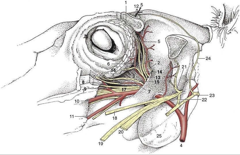
Figure 18-36 Dissection of the orbit; the zygomatic arch and periorbita have been removed.
1, Lacrimal gland; 2, periorbita; 3, lateral rectus; 4, maxillary a.; 5, supraorbital a.; 6, lacrimal a.; 7, muscular branch of external ophthalmic a.; 8, malar a.; 9, infraorbital a.; 10, major palatine a.; 11, buccal a.; 12, supraorbital n.; 13, lacrimal n.; 14, trochlear n.; 15, zygomatic n.; 16, oculomotor n.; 17, rostral branches of maxillary n.; 18, buccal n.; 19, lingual n.; 20, inferior alveolar n.; 21, masticatory n.; 22, auriculotemporal n.; 23, facial n.; 24, auriculopalpebral n.; 25, guttural pouch.lateral aspect of the bulbus, where it is protected by the adjacent part of the orbital rim (Figure 18—36/7). A small accessory lacrimal gland is associated with the deep part of the cartilage of the third eyelid.
The nasolacrimal duct, already mentioned in relation to surgical access to the maxillary sinus, provides a conspicuous feature where it opens on the floor of the nostril (see Figure 18-3). The extraocular muscles show little that is distinctive; as is common in ungulates, the retractor bulbi is relatively large (see Figure 9-19/7).
The eyeball shows significant departure from the spheroidal form—it is compressed from front to back and is higher than it is wide—which is relevant to the concept of the ramp retina (see further on). It is constructed of the usual layers. The sclera is relatively thin toward the equator, where it obtains a bluish tint from the pigmentation of the underlying choroid. The cornea is relatively small and ovoid; its pointed end is lateral.
The choroid exhibits a triangular green or bluish- green tapetum dorsal to the optic disc (Figure 18-37). The ciliary muscle is poorly developed; a second point is adduced in support of the theory of the ramp retina as the means of accommodation. The iris is generally
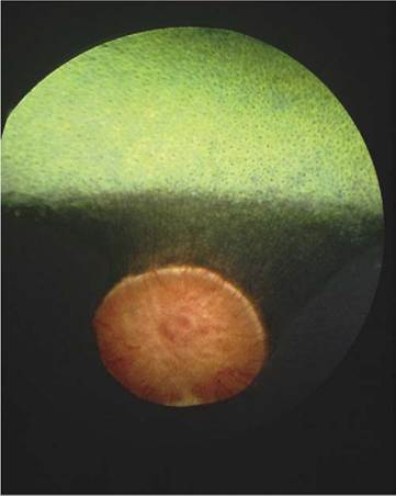
Figure 18-37 Fundus of eye of horse.
dark brown; in the absence of pigmentation (a rather uncommon anomaly) it is a rather unattractive bluish color (“walleye”) (see Figure 9-10, B). Both the iris and the pupillary opening within it are oval (with the long axes horizontal), but the pupil becomes rounder when contracted. The pupil of the newborn is almost round. Both margins of the pupil, but particularly the upper one, carry irregular granular excrescences interpreted as “shades” that limit the entry of light (see Figure 9-9/5).
The optic disc, very prominent on ophthalmoscopic examination of the fundus, is placed ventral to the tapetum and ventrolateral to the posterior pole of the bulb (see Figure 18-37). The macula is said to comprise both round and elongated parts; it is asserted that the former is concerned with binocular vision, the latter with monocular vision. The central artery of the retina is poorly developed, and the few straight branches that radiate from the margins of the disc soon fade. Much the larger part of the retina is nourished by the vessels of the middle tunic. There is nothing noteworthy in the refractive media.
It is believed that the poor development of the ciliary muscle compels the horse to rely on the distorted form of the bulb for accommodation. The upper part of the
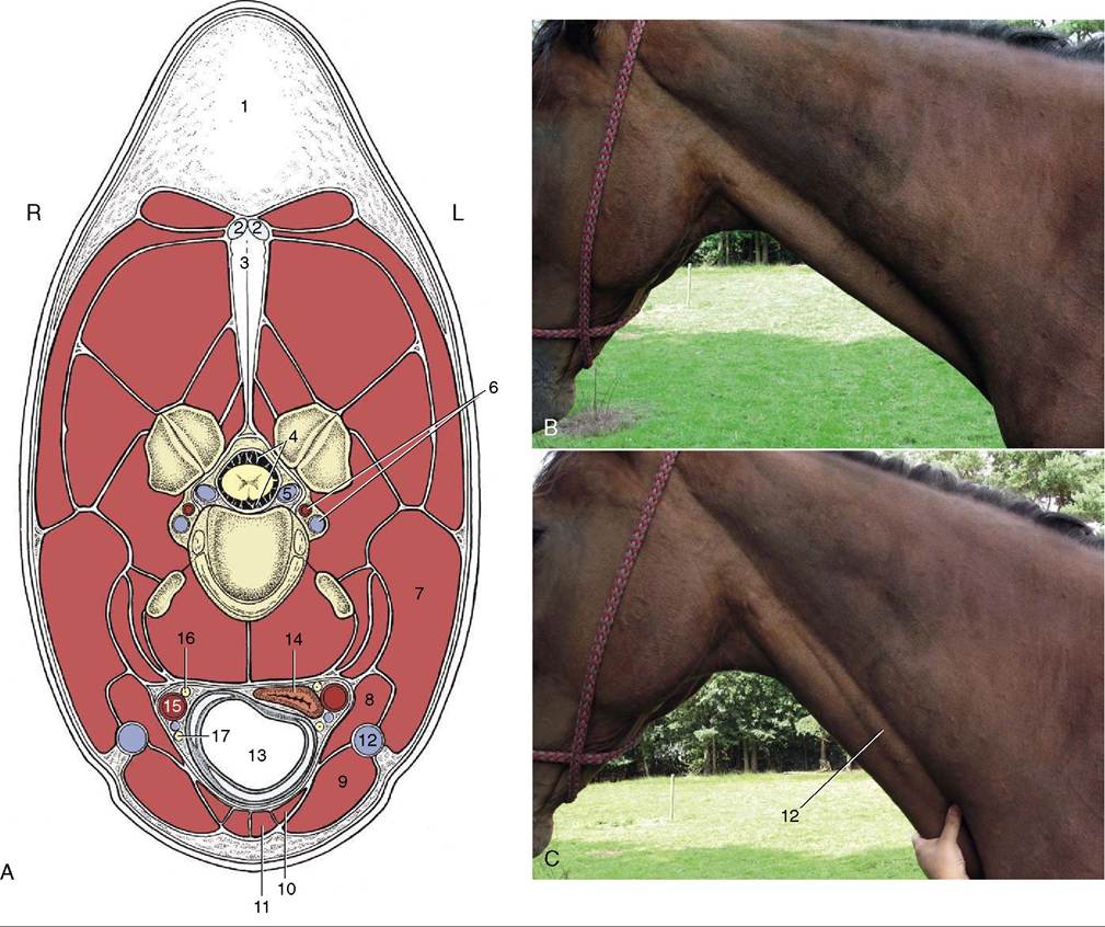
Figure 18-38 A, Transection of the neck at the level of the fourth cervical vertebra. B, The external jugular vein is not visible, but it is raised (C) when occluded in the jugular groove. 1, Crest; 2, 3, funicular and laminar parts of nuchal ligament; 4, subarachnoid space; 5, internal vertebral venous plexus; 6, vertebral artery and vein; 7, brachiocephalicus; 8, omohyoideus; 9, sternocephalicus; 10, sternothyroideus; 11, sternohyoideus; 12, external jugular vein; 13, trachea; 14, esophagus; 15, common carotid artery; 16, vagosympathetic trunk; 17, recurrent laryngeal nerve.
retina, which is at a greater distance from the lens, serves for near vision; the lower part, closer to the lens, serves for distance vision. The animal therefore adjusts the carriage of the head—and thereby the location of the image on the retina—as a means of focusing. The technique is sometimes well-illustrated by a horse approaching and jumping an obstacle.