THE EYEBALL
The eyeball (bulbus oculi) of the domestic mammals is nearly spherical but with some anteroposterior compression in horses and cattle. In addition, the cornea, the transparent part of the eyeball, bulges from the anterior surface by virtue of its smaller radius of curvature (Figure 9-2).
The highest point on the cornea is the anterior pole, and the highest point on the posterior surface is the posterior pole of the eyeball; the straight line passing through both poles is the optic axis. The equator is an imaginary line about the eyeball, which, like that of the Earth, is equidistant from the poles. A meridian is one of the many lines passing from pole to pole that intersect the equator at right angles. The optic nerve (Figure 9-2/6) leaves the eyeball slightly ventral to the posterior pole.
The eyeball has three thin tunics that, being in close apposition, form a laminated sheet that surrounds the partly liquid, partly gelatinous center. The three tunics are (1) an external fibrous tunic that gives form to and protects the eyeball—it is the only complete tunic; (2) a middle vascular tunic that consists largely of blood vessels and smooth muscle and is concerned with the nutrition of the eyeball and the regulation of the shape of the lens and size of the pupil; and (3) an internal nervous tunic that consists largely of nervous tissue and is the layer most directly concerned with vision, that is, the translation of visual stimuli into nerve impulses for interpretation by the brain.
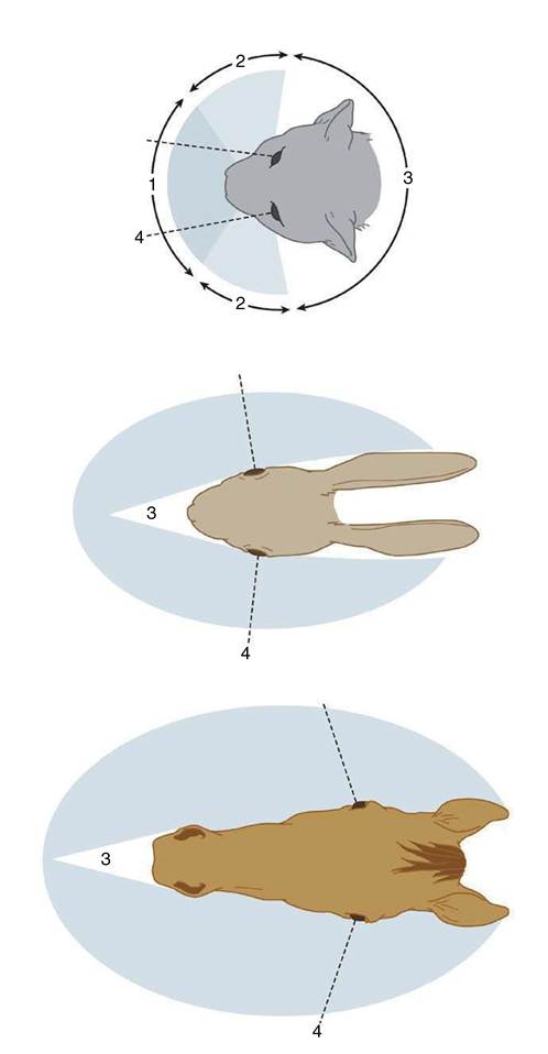
Figure 9-1 Visual fields of cat, rabbit, and horse. 1, Binocular vision; 2, monocular vision; 3, blind area; 4, visual axis of eye in central position.
The Fibrous Tunic
The fibrous tunic of the eyeball is made up of very dense collagenous tissue, which, by resisting the internal pressure, gives the eye shape and stiffness.
It consists of the sclera and cornea, which meet at the limbus (Figure 9-2/7).The sclera is the opaque posterior part of the fibrous tunic. It consists of a dense feltwork of collagenous and elastic fibers and is generally white (“the white of the eye”), though with a bluish tinge; in some species it contains pigmented cells that render it gray. Ventral to
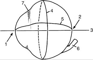
Figure 9-2 Medial view of right eyeball. 1, Anterior pole; 2, posterior pole; 3, optic axis; 4, equator; 5, a meridian; 6, optic nerve; 7, limbus.
the posterior pole it presents a small cribriform area (Figure 9-3/13) through which pass the fibers of the optic nerve. The nerve is surrounded by a connective tissue sheath that continues the dura mater to the sclera. The sclera is also pierced by several small ciliary arteries and nerves and by larger vorticose veins. It gives attachment to the tendons of the ocular muscles anterior to the equator. Posteriorly, except for the areas taken up by the retractor bulbi muscle, it is covered by a thin membrane (vagina bulbi; Figure 9-3/5) that separates it from the retrobulbar fat, which provides a socket in which the eyeball can play. Near the limbus the sclera is covered by conjunctiva (see further on), which furnishes connection to the inside of the lids (Figure 9-3/19).
The cornea forms about one quarter of the fibrous tunic and bulges forward (Figure 9-4). It is composed of a special kind of dense connective tissue arranged in lamellar form. It is generally recognized that, despite the careful arrangement of its fibers, transparency is not only a structural but also a physiological phenomenon and depends on the continuous pumping out of interstitial fluids, which is a process that has been localized in the posterior epithelium. Its main bulk (substantia propria) is continuous with the sclera (Figure 9-5/6,9) and encased by anterior and posterior limiting membranes and epithelial layers.
The anterior epithelial layer is continuous with the epithelium of the conjunctiva, while the posterior epithelial layer unites with the anterior surface of the iris across the iridocorneal angle (Figure 9-5/4). The cornea does not contain blood vessels; nutrients for its cells permeate the substantia propria from vessels in the limbus or are carried to its surfaces in the lacrimal fluid and aqueous humor. The surface of the cornea is very sensitive owing to the presence of free nerve endings near the anterior epithelium. These arise from the long ciliary nerves, which are branches of the ophthalmic nerve (see further on). Their axons form the afferent limb of the corneal reflex, which closes the lids when the cornea is touched. This reflex is employed when monitoring deep anesthesia.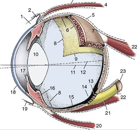
Figure 9-3 Eye opened to show the three tunics, which have been drawn thicker than they actually are. 1, Limbus; 2, upper fornix; 3, deep muscular fascia; 4, dorsal rectus muscle; 5, vagina bulbi; 6, choroid; 7, sclera; 8, ora serrata; 9, retina; 10, lens; 11, optic axis; 12, visual axis; 13, area cribrosa; 14, optic disc; 15, retina; 16, ciliary body; 17, iris; 18, cornea; 19, conjunctiva; 20, ventral rectus muscle; 21, optic nerve; 22, retractor bulbi; 23, sheath of optic nerve.
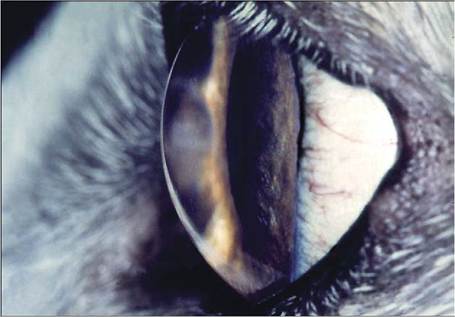
Figure 9-4 Curvature of canine cornea.
The Vascular Tunic
The vascular tunic of the eye (also known as the uvea) lies deep to the sclera to which it is applied. It consists of three zones: choroid, ciliary body, and iris, given in posteroanterior sequence (see Figure 9-3). The choroid lines the sclera from the optic nerve almost to the limbus, the ciliary body follows as a thickened zone opposite the limbus, and the iris projects into the cavity of the eyeball posterior to the cornea; the iris is the only
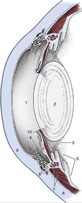
Figure 9-5 Anterior part of the eye in section.
1, Anterior chamber; 2, lens; 3, zonular fibers; 4, iridocorneal angle; 5, ciliary body; 6, sclera; 7, ciliary muscles; 8, venous plexus of sclera; 9, cornea; 10, iris with the sphincter and dilator muscles shown.internal structure readily seen through the cornea without recourse to instruments (ophthalmoscope). Although blood supply is its principal function, the vascular tunic suspends the lens, regulates its curvature, and adjusts the size of the pupil by means of the smooth muscle in the ciliary body and iris (see Figure 9-5).
The choroid contains a dense network of blood vessels embedded in heavily pigmented connective tissue. The network is supplied by the posterior ciliary arteries and is drained by the vorticose veins. A flat sheet of capillaries on the internal surface is responsible for the nutrition of the external layers of the nervous tunic (retina), which lies deep (internal) to it. The blood in these capillaries produces the redness of the fundus (interior surface of the posterior hemisphere) seen when the eye is examined with an ophthalmoscope. In the dorsal part of the fundus the choroid forms a variously colored, light-reflecting area known as the tapetum lucidum (Figure 9-6, A-F). This is an avascular layer (cellular in carnivores, fibrous in ruminants and horses) between the capillaries and the network of larger vessels. The tapetal cells contain crystalline rods arranged in such a way that light striking them is split into its components, which results in the characteristic iridescence. The packaging of the collagen in the fibrous tapetum has the same effect. The tapetum makes the eyes of animals “shine” when they look toward a light, such as the headlights of an oncoming car. Our eyes, and those of the pig, do not have a tapetum and therefore do not give this effect. It is believed that the tapetum is a nocturnal adaptation: by reflecting incident light, it increases the stimulation of the light-sensitive receptor cells in the overlying retina and thus aids vision in dark places.
The choroid adheres so closely to the pigmented external layer of the retina that the latter remains when the bulk of the retina is removed during dissection. The retina is without pigment where it overlies the tapetum lucidum.Toward the limbus the choroid thickens to form the ciliary body (Figure 9-5/5). This is a raised ring with ridges converging toward the lens in the center; anteriorly the ring is continued by the iris. The ciliary body is best comprehended when seen in its entirety by looking into the anterior part of the eye from behind (Figure 9-7/2; Figure 9-8). The radial ridges, known as the ciliary processes, extend zonular fibers (Figure 9-5/5) to the equator of the lens, suspending it around its periphery. Between the ciliary body and the sclera is the smooth ciliary muscle (Figure 9-5/7), which functions in accommodation (the ability of the eye to focus on near or distant objects by changing the shape of the lens) (see further on).
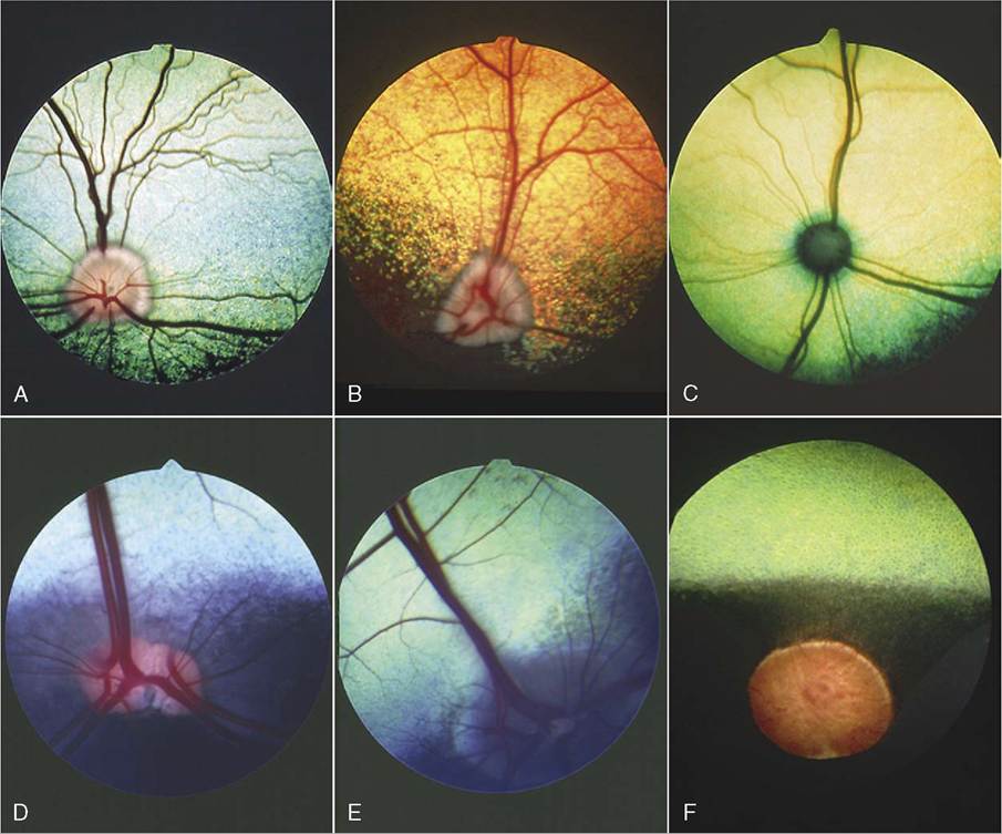
Figure 9-6 A to F, Fundus of eye. A, Dutch sheepdog. B, Old English sheepdog. C, Cat. D, Cow. E, Goat. F, Horse.
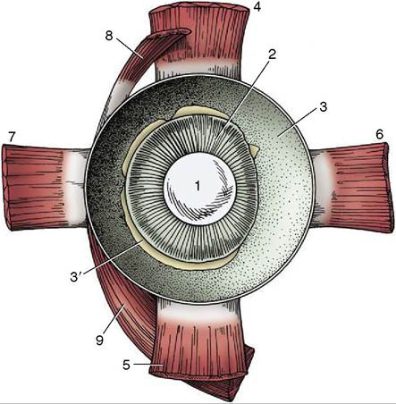
Figure 9-7 Anterior half of the left equine eye, viewed from behind. 1, Lens; 2, ciliary body; 3, choroid covered by pigmented outer layer of retina; 3', remnants of inner nervous layer of retina, which has been removed; 4-7, dorsal, ventral, medial, and lateral rectus muscles; 8,9, dorsal and ventral oblique muscles.
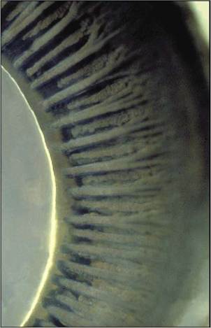
Figure 9-8 Posterior view of ciliary body with ciliary processes (horse).
The third and smallest part of the vascular tunic is the iris (Figure 9-5/10), which is suspended between the cornea and lens. It is a flat ring of tissue attached at its periphery to the sclera (by the pectinate ligament; Figure 9-12/7) and to the ciliary body; the opening in
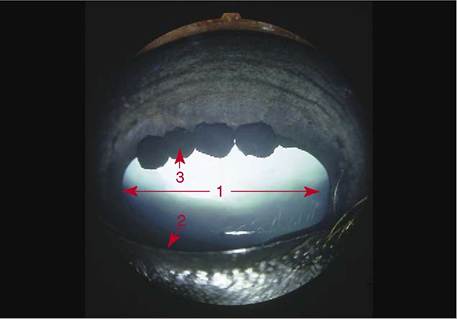
Figure 9-9 Anterior surface of the equine iris with characteristic iridic granules.
1, Pupil; 2, pupillary margin; 3, iridic granule.the center is the pupil (Figure 9-9) through which light enters the posterior part of the eye. The size of the pupil and therefore the amount of light reaching the retina are regulated by smooth sphincter and dilator muscles in the iris. The sphincter lies near the pupillary margin, while the fibers of the dilator are arranged radially and, on contraction, enlarge the pupil. Irregular outgrowths (iridic granules; Figure 9-9) containing coils of capillaries are often seen on the upper and lower pupillary margins of ungulates; their significance is not known, though there are suggestions that they act as “shades.”
The iris divides the space between the lens and cornea into anterior and posterior chambers that communicate through the pupil (see Figure 9-9). Both are filled with aqueous humor, a clear watery fluid (see further on).
The iris consists of three layers: an anterior epithelial layer continues across the iridocorneal angle and blends with the posterior epithelium of the cornea, a middle layer of connective tissue stroma contains the two smooth muscles, and the posterior layer of pigmented epithelium is the forward extension of the pigmented layer of the retina mentioned when we described the choroid; it is known as the iridic part of the retina and is closely related to the dilator pupillae (Figure 9-5/10).
The color of the iris determines the “color of the eye” and depends on the number of pigmented cells present in its stroma and on the type of pigment in the cells. If the pigmented (melanin) cells are tightly packed the iris is dark brown, with fewer cells the iris is lighter and yellowish (Figure 9-10), and with a minimum of pigmented cells the iris appears bluish. In albinos pigment is also absent from the iridic part of the retina, that is, the iris is totally devoid of pigment; their eyes appear red because the blood in the capillaries is not obscured.
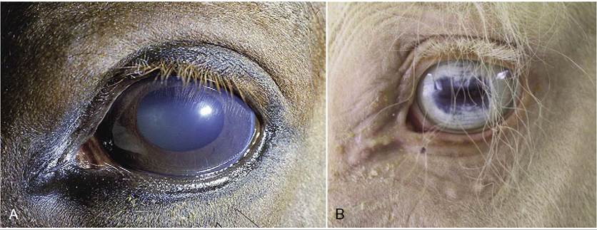
Figure 9-10 A, Left equine eye. Note the brown pigmentation of the iris. B, Left equine eye of an albino animal. Note the absence of pigment.
The Internal Tunic
The internal or nervous tunic of the eyeball contains the light-sensitive receptor cells and is known as the retina (Figure 9-3/9,15). It is an extension of the brain to which it remains connected by the optic nerve. The retina begins where the nerve penetrates the choroid; shaped like a hollow cup, it lines this and ends at the pupillary margin. Only the posterior two thirds or so of the retina can be reached by light entering the pupil. Consequently, only that part (pars optica retinae) is provided with receptor cells; it is relatively thick. The remaining third is “blind” (pars ceca retinae) and is mainly represented by the thin pigmented layer that continues on to the ciliary body and the back of the iris. The edge caused by the abrupt decrease in thickness at the junction of optic and blind parts is the ora serrata (Figure 9-3/8); it also demarcates the choroid from the ciliary body. The two layers of the retina develop from the inner and outer layers of the optic cup with which the eye makes its appearance in the embryo. The gap between the layers of the optic cup, though obliterated postnatally, remains a weakness where delamination produces “detachment of the retina.”
The presence of so much retinal and choroidal pigment makes the interior of the posterior part of the eye dark like the inside of a camera so that the pupil appears black. The black walls absorb scattered and reflected light and prevent it from striking the retina a second time, which would contribute to blurred vision.
The layers in the pars optica retinae are as follows, beginning at the choroid: a single layer of pigmented cells; a neuroepithelial layer containing the receptor cells, rods and cones and their nuclei (the rods, so far as we know, are concerned with black and white [night] and the cones with color [day] vision); a layer of bipolar ganglion cells; and a layer of multipolar ganglion cells whose nonmyelinated axons, lying internal (deep) to the cells, pass to the optic disc, where they aggregate to form the optic nerve. It will be clear from this arrangement that light passes through all layers except the first before reaching and stimulating the rods and cones (Figure 9-11).
The area where the axons of the fourth layer concentrate to leave the eye, the optic disc, can easily be seen when examining the fundus with an ophthalmoscope (see Figure 9-6). Because the axons here turn in toward the cribriform area of the sclera, there is no room for receptor cells; the optic disc, therefore, is a blind spot. In contrast, an area of maximum optical resolution (macula) is located a short distance dorsolateral to the optic disc. It is believed that when we examine objects intently, we focus them on the macula. It is not known whether animals do the same. In some species the macula is faintly visible with the ophthalmoscope. The visual axis is the line connecting the macula, the center of the lens, and the object viewed. It does not quite coincide with the optic axis because the macula is slightly dorsal to the posterior pole of the eyeball (see Figure 9-3).
Arterioles and venules emerging from the optic disc spread out in various species-specific patterns to nourish and drain the retina (see Figure 9-6). The arterioles are branches of the central artery of the retina, which arrives at the optic disc in the center of the optic nerve.
The anteroposterior compression of the equine eyeball has led to the assumption that the horse has a ramp retina. A ramp retina is one in which all parts are not equidistant from the posterior pole of the lens; the distance from the lens becomes progressively greater as the retina is followed dorsally. Presumably, as increasingly closer objects are viewed, they are focused on the more dorsal parts of the retina; focal length is automati-
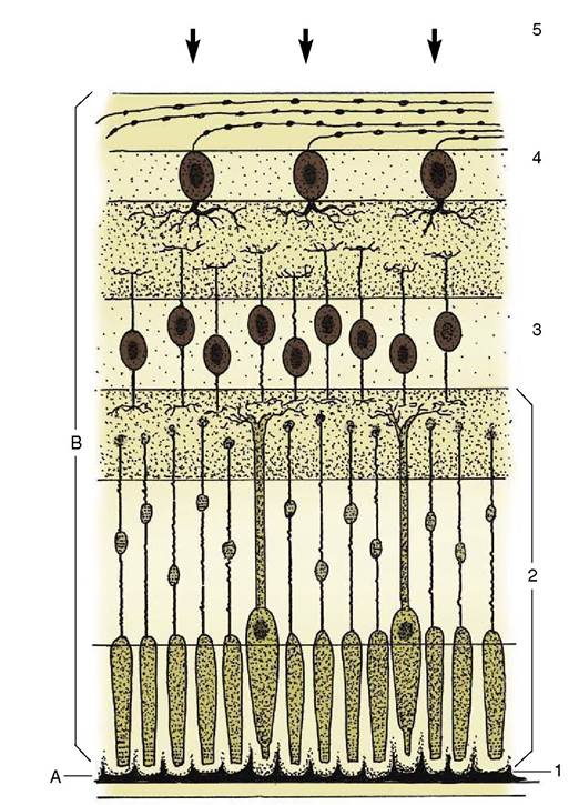
Figure 9-11 Outer pigmented layer (A) and inner neuroepithelial layer (B) of retina. 1, Pigmented cells; 2, receptor cells (rods and cones); 3, bipolar ganglion cells; 4, multipolar ganglion cells; 5, incoming light (arrows).
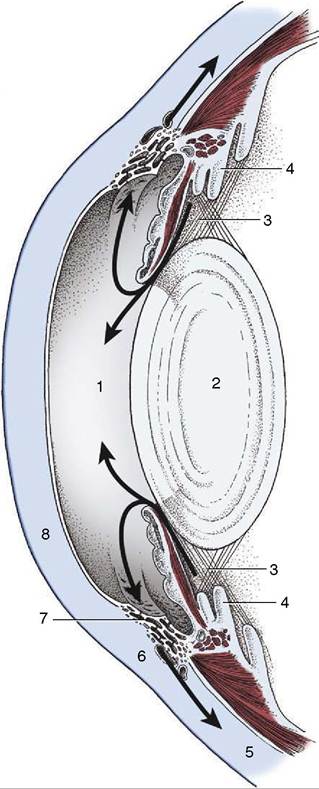
Figure 9-12 The flow (arrows) of aqueous humor. 1, Anterior chamber; 2, lens; 3, posterior chamber; 4, ciliary body; 5, sclera; 6, venous plexus; 7, pectinate ligament; 8, cornea.
cally increased, and little accommodation of the lens is required (p. 528).
The Refractive Media of the Eyeball
Now that the laminated wall has been described, it remains to say something of the interior of the eyeball, which is concerned with the manipulation of the light rays that enter it. It is best to do this by following the path taken by the light. Several interior structures have already been mentioned and require little further description.
The cornea is an integral part of the supporting fibrous tunic. Although dense and tough, it has the quality of being transparent and thus enables light to enter the eye. The cornea plays a major role in refraction; that is, it is capable, as is the lens, of bending light so that what is seen by the animal is miniaturized sufficiently to be focused on the retina.
The rays next encounter the aqueous humor filling the space between cornea and lens. The aqueous humor is a clear watery fluid that, apart from its refractive properties, plays an important role in the maintenance of intraocular pressure. It is continuously produced by cells of the ciliary processes and enters the system in the posterior chamber. From here it passes through the pupil into the anterior chamber and thence through the spaces in the trabecular tissue (pectinate ligament) at the iridocorneal angle. These spaces convey it to venous sinuses in the sclera and thus into the bloodstream (Figure 9-12). In the healthy eye the rate of production balances the rate of drainage, maintaining a constant pressure. Interference with drainage allows excess fluid to accumulate, causing the intraocular pressure to rise (glaucoma). This serious condition, common in humans, is rarely seen in animals.
The lens (Figure 9-13), in contrast to its liquid neighbors, is a solid structure, though sufficiently elastic to be able to change in shape. It is biconvex and has anterior and posterior poles, an equator, and a central axis that coincides with the optic axis of the eye. The posterior surface is usually more convex than the anterior. The lens has an outer capsule that is thicker anteriorly and thickest at the equator, where the zonular fibers of the ciliary body are secured. The capsule of the lens is elastic and is permanently under tension, which, if unopposed by the pull exerted at the periphery, would cause the lens to assume a more spherical shape. The substance of the lens consists of very regularly arranged fibers. These form concentric sheets that can be peeled
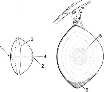
Figure 9-13 Bovine lens; on the right, a meridional section. 1, Anterior pole with lens star; 2, posterior pole with lens star; 3, equator; 4, optic axis; 5, nucleus; 6, layers of lens fibers, shown only in part.
off like the layers of an onion. Within each sheet the fibers are so arranged that they loop from a point on the anterior surface to one on the posterior surface. Their ends are cemented to the ends of other fibers, forming visible sutures shaped like little three-pointed stars (radii lentis; Figure 9-13/1,2). In the peripheral, or cortical, part of the lens the fibers are relatively soft; they are firmer and thinner toward the center where they form a harder nucleus. Owing to its elastic properties the cortex can be molded so that the lens changes shape during accommodation. In many older animals the lens becomes cloudy, impairing vision; the condition is known as cataract (Figure 9-14).
Accommodation. As we have said, the elastic capsule of the lens would squeeze the relatively soft cortex into a rounder shape unless opposed by the zonular fibers that arise from the ciliary processes and exert a constant radial pull on the equator. This pull flattens the lens into the shape described; this is the resting shape of the lens adapted for far vision and is present during sleep. When the animal wants to focus on a near object the muscle on the surface of the ciliary body contracts, thickening the ciliary body. This displaces the processes toward the lens and thus relaxes the zonular fibers. The lens, released from the tension at its equator, rounds out and brings the object into focus. Compared with the muscle in humans, the ciliary muscle and, therefore, the ability to accommodate are poorly developed in domestic animals.
After passing through the lens the light rays enter the vitreous body. This is a gel-like mass consisting mainly of water (vitreous humor) but with a stroma of fine transparent fibers that condenses into a membrane at the surface. The vitreous body occupies the space between lens and retina and holds the latter against the choroid. In the embryo the lens is nourished by the
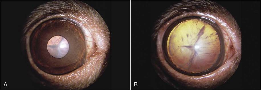
Figure 9-14 A, Slightly constricted canine pupil. Cataract of lens visible. B, Canine pupil in mydriasis (enlarged pupil): Lens is now totally visible; opacity is seen to affect the entire lens.
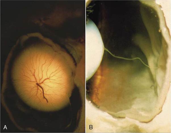
Figure 9-15 A, Posterior surface of lens (newborn puppy) showing remnant of hyaloid a. B, Persistent hyaloid a. (dog).
hyaloid artery, a branch of the central retinal artery that passes through the vitreous body. The artery usually degenerates after birth, and the lens is then nourished by diffusion (Figure 9-15, A-B). Unlike the aqueous humor, the vitreous humor is not continuously replaced; it is therefore constant in volume.