The Eye's Anatomy Is Adapted to the Eye's Role as a Visual Receptor Organ
Figure 14-1 shows the anatomy of the normal eye in the horizontal plane. The white, outer protective layer encasing most of the eyeball is called the sclera. It is modified anteriorly into a clear, stratified squamous epithelial layer called the cornea.
In the posterior two thirds of the eye the sclera is lined with a vascular and pigmented layer called the choroid.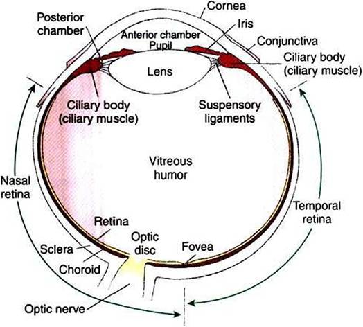
FIGURE 14-1 Schematic diagram of a horizontal section through the right eye as viewed from above. (Redrawn from Walls GLiThe vertebrate eye and its adaptive radiation, Cranbrook Institute of Science, Bulletin 19, 1942.)
Interior to the choroid is the retina, the layer containing the photoreceptors.
As light passes through the transparent cornea» it undergoes some bending that will ultimately help to focus it on the retina. After passing through the cornea» the light enters a compartment called the anterior chamber. The anterior chamber and the posterior chamber are filled with a clear, waterlike fluid called aqueous humor that supplies important nutrients to the cornea (as well as the lens). Separating the anterior and posterior chambers is a diaphragm of varying size called the iris. The iris is a pigmented structure containing dilator and constrictor smooth muscle fibers arranged to vary the diameter of the pupil, the hole in the iris through which light passes on its way to the retina. The size of the pupil regulates the amount of light entering the eye. Behind the iris is the lens. The lens is suspended in the eye by the suspensory ligaments, which attach to the lens and to the ciliary body, a muscular structure at the base of the iris. The lens provides variable focusing power, in contrast to the fixed cornea.
Behind the lens is a chamber filled with a gelatinous fluid called the vitreous humor.
Because of the viscosity of this fluid, the pressure generated by the aqueous humor, and the fairly inelastic nature of the sclera and cornea, the globe of the eye is basically spherical. The vitreous humor can also clear ocular debris potentially obstructing the light path. Behind the vitreous humor is the neural retinal layer where light is transduced into the electrical activity of neurons. The retina is interrupted at a point where axons of the retina’s ganglion cell layer, which travel across the inner surface of the retina, leave the eye on their way to the brain. This point, the optic disc, is a recognizable structure when the eye is examined with an ophthalmoscope (Figure 14-2). The interruption of the retina at the optic disc produces a “blind spot,” another name for the optic disc. The retinal ganglion cell axons leaving the eye at the optic disc give rise to the optic nerve (cranial nerve II), a cranial nerve so rich in axons that there are more axons in both optic nerves than in all the dorsal roots of the spinal cord.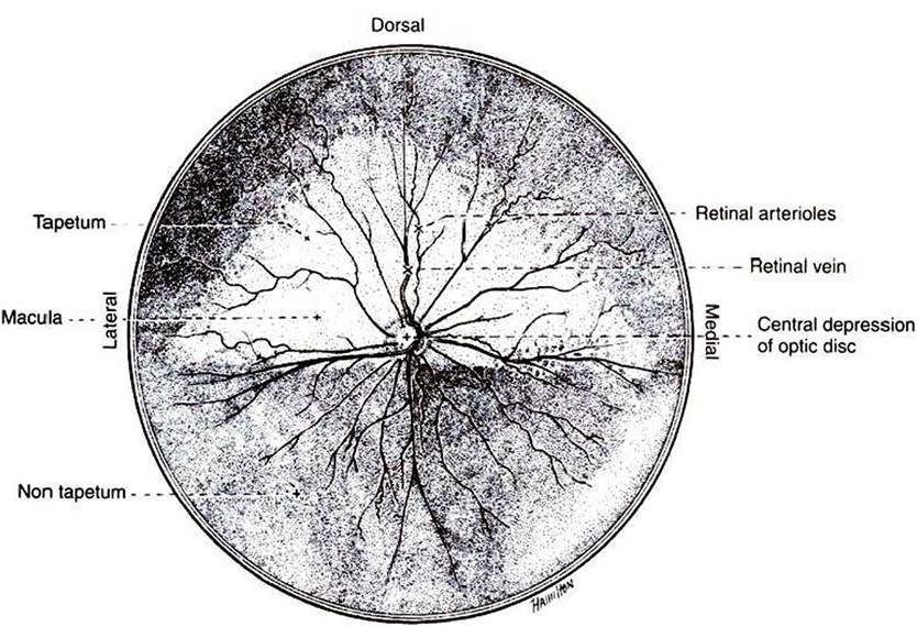
Ventral
FIGURE 14-2 Ocular fundus of the right eye of a dog, as viewed with an ophthalmoscope, showing the optic disc and retinal blood vessels. (From Evans HE, Christensen GC: Miller's anatomy of the dog, ed 2, Philadelphia, 1979, Saunders.)
Also visible through the ophthalmoscope, on the surface of the retina, are the retinal blood vessels (see Figure 14-2). I his network of arteries and veins enters the retina at the optic disc and provides much of the nutrition to the retina. Vessels of the choroid provide the remaining nutrition to the retina. Examination of retinal vessels often provides valuable clues to abnormalities elsewhere in the cardiovascular system.
The lacrimal gland, located near the lateral canthus of the eye, produces tears in response to parasympathetic nerve stimulation. Tears then flow over the cornea and are drained into the nose by the nasolacrimal duct. A regular flow of tears across the cornea is essential to the health of the cornea.