THE FOREBRAIN
The forebrain comprises the median diencephalon and the paired cerebral hemispheres (telencephalon). The hemispheres overlap the dorsolateral aspects of the diencephalon to which they have become fused by the growth of fiber tracts across the gaps.
The Diencephalon
The diencephalon (there is no convenient alternative name) forms the most rostral part of the brainstem. Only its most ventral part, the hypothalamus, is visible on the external surface of the intact brain (Figure 8-19), but it is more extensively revealed in median section (see Figure 8-22). The diencephalon comprises three parts: epithalamus, thalamus (including subthalamus), and hypothalamus, which develop in relation to the roof, walls, and floor of the third ventricle, respectively.
The epithalamus, the most dorsal part, comprises the pineal gland (epiphysis cerebri), habenular striae, habenulae, and habenular commissure (Figure 8-30). The pineal gland (Figure 8-30/d) is a small, median body projecting dorsally from the brainstem behind an evagi- nation of the roof of the third ventricle that is composed only of pia and ependyma. Although the pineal gland has long been suspected to play some part in sexual development and behavior, its functions are only now becoming clear; it is believed to be particularly concerned in the seasonal regulation of ovarian activity
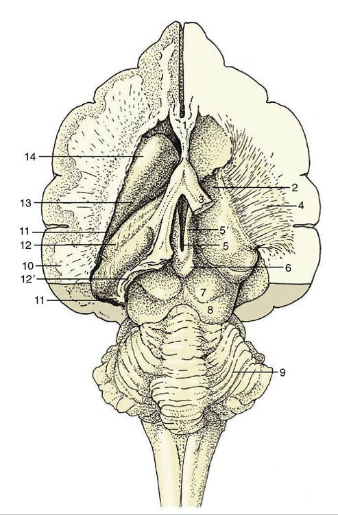
Figure 8-30 Dorsal view of the canine brain. Part of the left hemisphere has been removed, which opens the lateral ventricle. On the right, the hippocampus and basal nuclei have also been removed, which exposes the thalamus and the internal capsule. 1, Septal nuclei; 2, dorsal surface of thalamus; 3, fornix (cut); 4, internal capsule; 5, dorsal part of third ventricle; 5', habenular nuclei (in roof of third ventricle); 6, epiphysis; 7, rostral colliculus; 8, caudal colliculus; 9, cerebellum; 10, cut lateral wall of hemisphere; 11, lumen of lateral ventricle; 12, hippocampus; 12', cut-edge of denticulate gyrus; 13, tail of caudate nucleus; 14, head of caudate nucleus.
in response to changing day length. The pineal gland produces melatonin, the pineal antigonadotropin that is also important in circadian and seasonal rhythms (p. 218). The habenular stria is a fiber bundle that among others connects the septal area with the habenular nuclei (Figure 8-30Z5'). It is an important pathway in the limbic system. The habenulae are nuclear complexes of enigmatic function that develop within the most dorsal parts of the ventricular walls. They receive fibers (habenular stria) from the hippocampus and other parts of the telencephalon and send fibers to mesencephalic
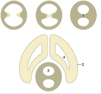
Figure 8-31 The formation of the interthalamic adhesion by median fusion of outgrowths of the lateral walls of the diencephalon. 1, Interthalamic adhesion; 2, telencephalon; 3, third ventricle; 4, lateral ventricle.
nuclei. The left and right habenular nuclei are interconnected via the habenular commissure.
The thalamus is the largest component of the diencephalon. It develops within the lateral walls of the third ventricle, but in many species, including domestic ones, it later bulges into the ventricle to form a bridge with its fellow. This, the intermediate mass or interthalamic adhesion, reduces the ventricle to an encircling annular space (Figure 8-31Z5). The relations of the thalamus are difficult to envisage because of its deep position and lack of separation from neighboring structures. It extends to the lamina terminalis grisea rostrally and to the midbrain caudally. Its dorsal surface faces toward the fornix and floor of the lateral ventricle, its ventral surface rests on the hypothalamus, and its lateral face is covered by the internal capsule of fibers ascending to and descending from the cerebral cortex (see Figure 8-30).
The thalamus is composed of a very large number of nuclei named according to their topographical relationships to each other. These nuclei have various specific functions and collectively form one of the most important relay and integration centers of the brainstem.
The ventral group receives most afferent systems (excluding the pathways concerned with olfaction) and also provides relays on feedback control systems of motor pathways (Figure 8-33).The subthalamus contains the subthalamic and endopeduncular nuclei and the zona incerta. The subthalamic nucleus acts as a relay station on the extrapy- ramidal motor pathway, whereas the other nuclei serve
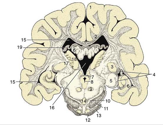
Figure 8-32 Transverse section of the canine brain at the boundary between the mesencephalon and diencephalon. 1, Cerebral hemisphere; 2, corpus callosum; 3, lateral geniculate nucleus; 4, optic tract; 5, medial geniculate nucleus; 6, hippocampus; 7, caudal commissure; 8, mesencephalic aqueduct; 9, red nucleus; 10, substantia nigra; 11, crus cerebri; 12, rostral extension of pontine nuclei; 13, middle cerebellar peduncle; 14, interpeduncular nucleus; 15, lateral ventricle; 16, third ventricle; 17, internal capsule; 18, thalamic nuclei; 19, fornix.
as links between the limbic system and the somatic and visceral motor systems.
The metathalamus, the Caudolateral part of the thalamus, comprises the medial and lateral geniculate bodies (Figure 8-32/5,5), whose presence and position were noted in the description of the midbrain. The lateral geniculate body, although not conspicuous in itself, is joined by the optic tract, which sweeps caudodorsally toward it, over the surface of the thalamus. The medial geniculate body lies ventromedial to the lateral one and receives acoustic fibers via the caudal colliculus (p. 300). The nuclei within these swellings relay visual and acoustic information to the cerebral cortex.
The hypothalamus forms the lower parts of the lateral walls of the third ventricle. It appears on the external surface of the brain between the preoptic region (rostral to the optic chiasm) and the cerebral peduncles and interpeduncular fossa (see Figure 8-19). Its salient surface features are the region known as the tuber cinereum, which extends the stalk or infundibulum that suspends the hypophysis below the brain, and the rounded mamillary body (see Figure 8-22) that receives information from the hippocampal complex and sends information to the thalamus (mammillothalamic tract of Vicq d’Azyr).
As such it is an important structure for memory. Internally it contains a number of nuclei associated with the visceral nervous system and hormonal regulation.The gonadotropin-releasing hormone (GnRH)- producing neurons have a curious history. They originate from outside the brain in the olfactory placode and migrate along the route taken by the developing olfactory, vomeronasal, and terminal nerves to enter the forebrain. Pheromone stimuli can directly influence the GnRH cells (see p. 352).
The hypophysis is a dark, solid body. It is located within a recess of the floor of the cranial cavity and is usually left behind when the brain is removed because the infundibulum, hollowed by a recess of the third ventricle, is easily torn across. The hypophysis is also held in place by a fold of dura mater (p. 308). The functions of the hypophysis are described elsewhere (p. 217).
The Telencephalon (Cerebrum)
The telencephalon consists of the paired hemispheres and the lamina terminalis grisea, the thin plate forming the rostral wall of the third ventricle with the organon vasculosum laminae terminalis griseae (Figure 8-66/7). Because the hemispheres develop as outgrowths of the diencephalon, their walls and lumina (lateral ventricles) remain in direct continuity with the corresponding features of that part. The adult hemispheres are semiovoid structures that form the largest part of the brain; their growth causes them to extend caudally over the brainstem to reach to within a short distance of the cerebellum. This growth brings them close together, and their flattened medial surfaces face toward each other across the narrow longitudinalfissure into which the falx cerebri fits when the brain is in situ. The remainder of the outer wall is divided between convex dorsolateral and flattish ventral (basal) surfaces (see Figures 8-20, 8-32, and 8-33).
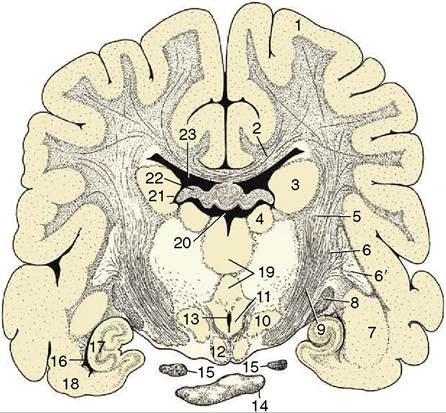
Figure 8-33 Transverse section of the canine brain at the transition between crus cerebri and internal capsule.
1, Cerebral hemisphere; 2, corpus callosum; 3, caudate nucleus; 4, thalamic nuclei; 5, internal capsule; 6, 6', lentiform nucleus; 6, globus pallidus; 6', putamen; 7, amygdala; 8, optic tract; 9, crus cerebri; 10, hypothalamic nuclei; 11, mammillotha- lamic tract; 12, mamillary body; 13, ventral part of third ventricle; 14, hypophysis; 15, oculomotor nerve; 16, ventral part of lateral ventricle; 17, hippocampus; 18, piriform lobe; 19, interthalamic adhesion; 20, dorsal part of third ventricle; 21, interventricular foramen; 22, fornix; 23, lateral ventricle.The walls of the hemispheres thicken unequally. Much of the medial wall of each hemisphere remains particularly thin, and in fetal life a part rolls inward, invaginating the pia mater and blood vessels covered by the ependymal lining into the ventricle, where it develops into the choroid plexus (p. 310) associated with this cavity. This structure produces the cerebrospinal fluid. The ventrolateral (striatal) part of the wall becomes much thickened when a number of large nuclei, the basal nuclei, develop within it. The alternation of these nuclei with the fiber aggregations in which they are embedded lends this region a striated appearance when exposed by section (see Figure 8-33); it is therefore appropriately known as the corpus striatum. The remainder of the wall is initially known as the pallium, but when it acquires an external covering of gray substance, again by migration from the ependyma, it is more frequently termed the cortex, although this term strictly designates only the outer gray substance.
Three regions of the pallium (or cortex) are distinguished on the basis of evolutionary history, structure, and function. The paleopallium initially served a purely olfactory function; it has retained this association in the highly developed mammals. The archipallium was also initially concerned with olfaction, but unlike the paleopallium, it has largely lost this association. The youngest part, the neopallium, made a very modest initial appearance in vertebrate history but has undergone a spectacular enlargement in mammals, in which it is both the largest and the functionally dominant part of the mammalian telencephalon.
These parts are now described separately, but in a different order for convenience. First, it may be helpful to dispose of the concept of a rhinencephalon (“smell-brain”) of primary olfactory function. Although it is true that the telencephalon of lower vertebrates developed specifically in relation to this sense, many parts have since discarded their original function and acquired new roles. The term rhinencepha- lon therefore no longer describes the functions of these parts at all adequately, and because it is now used in many conflicting ways, there is little in favor of its retention.The Paleopallium
The paleopallium is confined to the basal part of the brain; it is separated from the neopallium by the rhinal sulcus (Figure 8-34/4) on the lateral surface and, although less clearly, from the archipallium medially. Its rostral extremity is provided by an appendage, the olfactory bulb (Figure 8-34/7), that fits into a recess of the ethmoid bone. The surface apposed to the bone is made shaggy by the entrance of the numerous filaments that together form the olfactory nerve (I); these arise from receptors within the nasal mucosa and pass through the many perforations in the cribriform plate of the ethmoid bone. In the bulb the olfactory stimuli are conveyed to second-stage neurons. The bulb is continued caudally by the common olfactory tract (Figure 8-19/2), which soon divides into medial and lateral divisions separated by a triangular area. The medial tract runs toward the medial aspect of the hemisphere (precommissural area), where the information is conveyed to third-stage neurons. Some of the continuing fibers terminate within certain cortical gyri; others pass through the narrow anterior commissure in the rostral wall of the third ventricle to reach the corresponding region of the opposite hemisphere. The lateral tract continues caudally to join the large piriform lobe (Figure 8-19/3), the most salient feature of the basal surface of the hemisphere; not all the fibers in this tract reach the piriform lobe, as some are precociously detached en route, mainly to the amygdaloid body.
The Basal Nuclei
The large nuclei known by this title lie dorsal to the paleopallium, where a number of them combine with the white substance to form the corpus striatum. The complex may have had its original importance in relation to olfaction but has now acquired additional func-
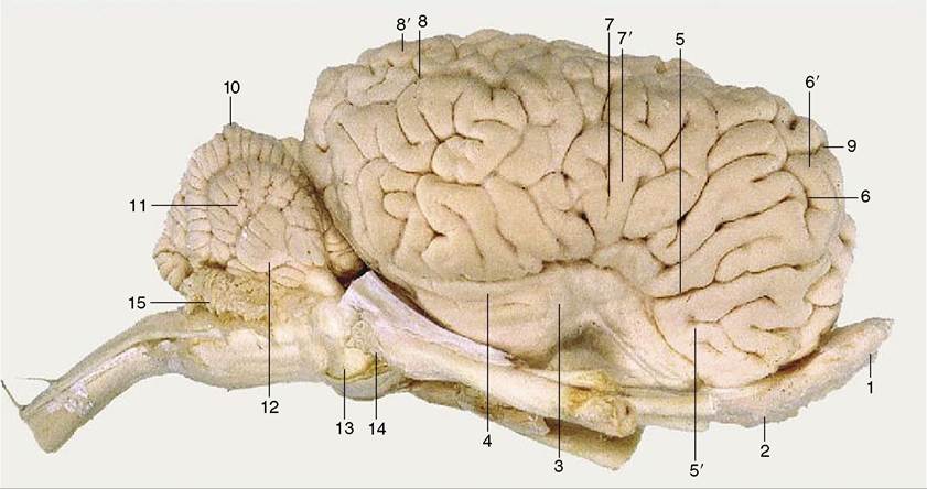
Figure 8-34 Lateral view of the equine brain. 1, Olfactory bulb; 2, olfactory tract; 3, piriform lobe; 4, rhinal sulcus; 5, sylvian sulcus; 5', sylvian gyrus; 6, ectosylvian sulcus; 6', ectosylvian gyrus; 7, suprasylvian sulcus; 7, suprasylvian gyrus; 8, ectomarginal sulcus; 8, ectomarginal gyrus; 9, cruciate sulcus; 10, cerebellar vermis; 11, cerebellar hemisphere; 12, paraflocculus; 13, pons; 14, crus cerebri; 15, caudal medullary velum.
tions in relation to other sensory input and to the regulation of motor function.
The nuclei composing the striatal complex are listed variously but most commonly as follows: the caudate nucleus, lentiform nucleus, amygdala, and claustrum. The caudate nucleus (Figure 8-33/5) has the general form of a comma with a large head bulging into the floor of the main part of the lateral ventricle, a body following the caudal bend of the cavity, and a tail related to the roof of its ventral extension (Figure 8-30/15,14). The Ientiform nucleus is more lateral and is divided by a fiber intersection into two parts: the medial globus pallidus and the lateral putamen (Figure 8-33/6,6'). The lentiform nucleus is separated from the caudate nucleus by the rostral limb of the fiber mass known as the internal capsule (Figure 8-33/5) and is separated from the thalamus by the caudal limb of the same formation. The nucleus accumbens, the reward center, is located in the ventral striatum.
The other basal nuclei are the smaller amygdala (Figure 8-33/7), located near the tail of the caudate nucleus, and the claustrum, which is interposed between the lentiform nucleus and neopallium. It is separated from these by other fiber laminae; the one on its lateral face is known as the external capsule.
The Neopallium
The neopallium constitutes the major part of the telencephalon: all that is visible in dorsal view and the bulk of that visible in lateral and medial views. References to the cortex, or even to the cerebrum without further qualification, usually have the neopallium specifically in mind. It is divided from the paleopallium by the rhinal sulcus on the lateral side of the hemisphere (Figure 8-21/4) and from the archipallium by the splenial sulcus medially (Figure 8-22/4). In some mammals, generally those that are of smaller size, its outer surface is smooth; however, in larger mammals, including domestic species, it displays a complicated arrangement of alternating ridges (gyri) and grooves (sulci) (see Figure 8-20). Though it is tempting to regard the more intricate modeling as evidence of greater intelligence and increased capacity for complex responses, the underlying cause appears to be physical. The ridges, which are mainly longitudinal, are produced by restraints imposed on the expanding telencephalic vesicle by the rigid corpus striatum and corpus callosum, while additional folding is necessary to maintain the relationship between volume (which increases by the cube) and cortical area (which increases by the square) in large brains.
The pattern of the gyri is reasonably constant within one species but differs among species. The features of greatest consistency include the cruciate sulcus, running transversely on the rostrodorsal aspect, a few sulci and gyri that follow the dorsomedial border, and the sylvian sulcus on the lateral side. Although other features provide useful landmarks for the investigator seeking to establish the functional significance of particular cortical areas, the names of most are of little consequence to the student. A simpler, rather arbitrary division of
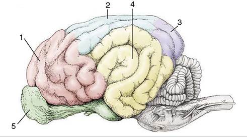
Figure 8-35 Cortical lobes of the canine brain. Lateral view. 1, Frontal lobe; 2, parietal lobe; 3, occipital lobe; 4, temporal lobe; 5, olfactory lobe.
more general utility distinguishes four regions or lobes named for their proximity to overlying bones; this division recognizes frontal, parietal, and occipital lobes in rostrocaudal sequence and a temporal lobe lying lateral to the last two. Only the frontal lobe is clearly demarcated because it is bounded caudally by the cruciate sulcus (see Figures 8-20/14 and 8-35).
The structure of the neopallium is more elaborate than that of other cortical areas and is remarkably uniform. It exhibits six superimposed strata that are densely populated by neurons and are separated by cell- free divisions. The neurons are broadly of two types: some more or less spherical (granular) neurons are provided with processes of very limited extent, and other (pyramidal) neurons have processes that range more distantly within the underlying white substance. The pyramidal neurons can be classified by their connections. Association fibers connect parts of the neopallium of the same hemisphere after passage directly below the cortex. Commissural fibers connect the two hemispheres, generally linking equivalent contralateral parts. They run over the roof of the lateral ventricle and mainly cross within the corpus callosum, the major tel- encephalic commissure that is shaped to form a rostral genu, middle trunk, and caudal splenium (Figure 8-22/3). Descending projection fibers from the cortex connect with lower parts of the central nervous system; most converge on the internal capsule squeezed between the basal nuclei and thalamus (Figures 8-36/7 and 8-37/1). In their courses to, from, and within the capsule the projection fibers are ordered according to their functional associations and somatotopic relationships.
The Archipallium
This part of the cortex was once concerned with the correlation of olfactory with other sensory information but has acquired new functions in modern mammals. It is included in the limbic system, which comprises the
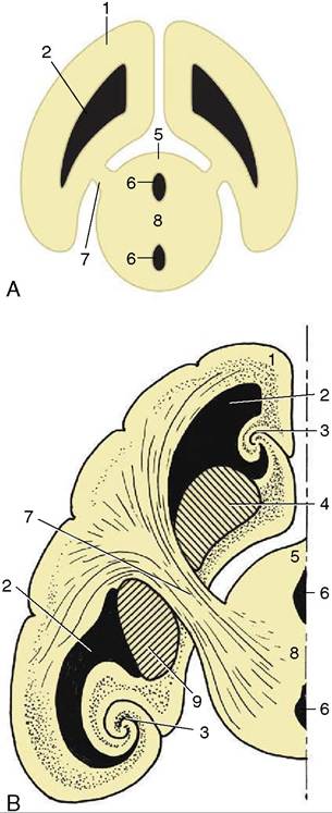
Figure 8-36 A, The connection between the cerebral hemisphere and diencephalon via the internal capsule (7). B, The lateral ventricle, basal nuclei, and hippocampus form concentric arches over the internal capsule. 1, Cerebral hemisphere; 2, lateral ventricle; 3, hippocampus; 4, caudate nucleus; 5, diencephalon; 6, third ventricle; 7, internal capsule; 8, interthalamic adhesion; 9, globus pallidus and putamen.
cingulate, supracallosal and geniculate gyri, the hippocampal formation, and the dentate gyrus.
The archipallium is no longer a conspicuous feature of the telencephalon. The relatively reduced importance of the olfactory sense and the enormous development of the neopallium have caused the archipallium to be displaced to the medial wall of the hemisphere; it is further reduced in prominence by a large part being rolled inward to lie on the floor of the lateral ventricle. The archipallium is topographically divided by the corpus callosum into a dorsal part that remains on the surface of the hemisphere (forming the cingulate and supracallosal gyri between the splenial sulcus and the corpus callosum; Figure 8-22/8,8') and a ventral part composed of the inflected portion usually known as the hippocampus (Figure 8-38/2). The archipallium is curved in conformity with the shape assumed by the expanding telencephalic vesicle and fits around the dorsal, caudal, and ventral aspects of the thalamus. This arrangement is difficult to envisage, and it is helpful to remember that the archipallium is interposed between
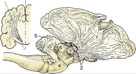
Figure 8-37 The internal capsule in the canine brain. A part of the cerebral cortex and the cortex of the cerebellum have been removed. The resected part of the telencephalon is indicated in the inset. 1, Fibers of the internal capsule; 2, optic tract, partly removed; 3, crus cerebri; 4, pons; 5, corpus medullare of cerebellum; 6, caudal colliculus; 7, medial geniculate body.
the olfactory bulb and the hypothalamus. The pathway is thus bent into a hairpin loop by the expansion of the hemisphere (Figure 8-39); the proximal limb extends, with a ventral concavity, caudally toward the apex of the loop, where a spiral twist sets the distal limb on a parallel returning course.
The proximal limb is provided by the surface gyri; beneath that run the longitudinal association fibers (cingulum) from the septal area. The fibers of this mul- tisynaptic pathway enter the caudal extremity of the hippocampus and form a covering to it. The fibers leaving the hippocampus run rostrally over its surface, gradually consolidating into a thick bundle, the fornix. The fornix lies directly below the corpus callosum at its commencement but deviates ventrally as it passes forward; it curves around the rostral extremity of the thalamus to enter the hypothalamus, where it terminates within the mamillary body (Figures 8-38 and 8-40). The right and left hippocampi are joined by the commissure of the fornix. There are thus three telencephalic commissures: the neopallial corpus callosum, the paleopallial anterior commissure, and the archipallial fornical commissure (also known as the commissure of the hippocampus).
When the fornix parts company with the corpus callosum, it remains connected to it by a thin septum that increases in depth toward its rostral end. This septum
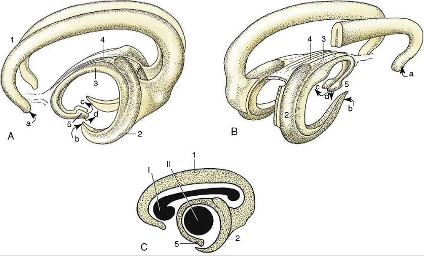
Figure 8-38 Three-dimensional representation of the archipallium. A, Left lateral view. B, Right caudolateral view. C, The positions of the corpus callosum (I) and the thalamus (II) are shown in lateral projection. 1, Supracallosal and cingulate gyri; 2, hippocampus; 3, fornix; 4, commissure of fornix; 5, hypothalamus with mamillary body. a, Input from the medial olfactory tract; b, input from the piriform lobe; c, output to the mammillothalamic tract; d, output to the brainstem.
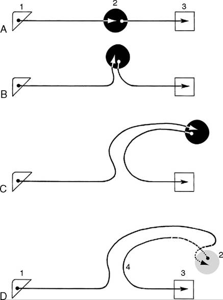
Figure 8-39 Diagram illustrating conjectured course of fibers running to and from the hippocampus. Because of differential growth of various parts of the brain, the hippocampus extends first dorsally (B), then caudally (C), and finally laterally (D). 1, Olfactory bulb; 2, hippocampus; 3, hypothalamus; 4, fornix.
telencephali (pellucidum) forms part of the medial wall of the lateral ventricle (Figure 8-22/28). It is a bilateral structure that is separated from its neighbor by a narrow, completely enclosed cleft and in its ventrorostral part contains septal nuclei in which fibers from the medial olfactory tract terminate.