THE MIDBRAIN
The midbrain (mesencephalon) is a short, rather constricted portion that better preserves the basic organization of the neural tube than do other parts of the brainstem.
The midbrain is exposed on the ventral surface of the intact brain, to which it contributes the crura cerebri, the interpeduncular fossa, and the superficial origin of the oculomotor nerves (III).
It is concealed dorsally by the overhanging cerebral hemispheres and cerebellum. Its lumen, the aqueduct, is a simple passage joining the much larger cavities of the third and fourth ventricles. The mesencephalon has a stratified structure, comprising tectum, tegmentum, ventral tegmentum and cerebral peduncle in dorsoventral sequence (Figure 8-29). Formally, all parts except the tectum are included within the cerebral peduncles, but in practice the latter term is frequently equated with the crus cerebri, the part ventral to the tegmentum.The tectum lies dorsal to the aqueduct. Its major features are four rounded surface swellings (see Figure
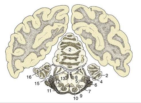
Figure 8-28 Transverse section of the canine brain at the level of the trigeminal nerve. 1, Cerebral hemisphere; 2, cerebellum; 3, rostral cerebellar peduncle; 4, lateral lemniscus; 5, rubrospinal tract; 6, root of V; 7, middle cerebellar peduncle; 8, medial longitudinal fasciculus; 9, medial lemniscus; 10, pyramidal tract; 11, pontine nuclei; 12, nucleus of lateral lemniscus; 13, reticular formation; 14, fourth ventricle; 15, rostral medullary velum; 16, root of IV.
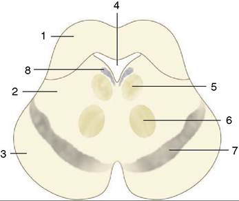
Figure 8-29 Schematic transverse section of the mesencephalon. 1, Tectum; 2, tegmentum; 3, crus cerebri; 4, mesencephalic aqueduct; 5, oculomotor nucleus (III); 6, red nucleus; 7, substantia nigra.
8, locus coeruleus.8-23). The paired caudal swellings, the caudal colliculi, are widely spaced and are joined by a substantial commissure. They are integration centers on auditory pathways (p. 300). There is a connection with the ipsilateral medial geniculate body (a swelling of the thalamus) via a distinct ridge (brachium). The rostral colliculi are placed closer together and are joined to the lateral geniculate bodies by similar but less obtrusive brachia. The rostral colliculi are staging posts on the visual pathways and are involved in somatic reflexes resulting from visual input, such as the response to being startled by a flash of intense light. They are also spatial integration centers.
The tegmentum comprises the core of the midbrain and is directly continuous with the corresponding stratum of the metencephalon. Much of it is formed by the reticular formation. The principal mesencephalic nuclei are the mesencephalic nuclei of the trigeminal nerves (V), the trochlear nuclei (IV), the principal and parasympathetic oculomotor nuclei (III), the red nuclei (named for their pronounced vascularity), and the periaqueductal gray, a core of gray substance about the aqueduct. The substantia nigra is a prominent lamina that can be identified in transverse sections by its darker color, which is due to the gradual accumulation of pigment within the constituent neurons. Like the red nucleus, it is associated with the basal nuclei (p. 291) in the control of voluntary movement.
The crura cerebri are visible on the ventral surface of the brain. They comprise fiber tracts that are in passage between the telencephalon and caudal brainstem. On emerging from the telencephalon, they converge, although they are separated by the interpeduncular fossa (Figure 8-19). The oculomotor nerves (III) emerge in this region, directly rostral to the pons.