The Fovea Solves a Distortion Problem Found in OtherAreas of the Retina
The retinal ganglion cells are located in the inner retina (closer to the vitreous humor), whereas the photoreceptor cells (rods and cones) are located in the outer retina (closer to the choroid; see Figure 14-4).
Therefore, throughout most of the retina, light rays travel through ganglion cells, bipolar cells, amacrine cells, and horizontal cells before reaching the photoreceptors. Although these inner neurons are unmyelinated (the ganglion cell axons become myelinated on leaving the eye) and therefore relatively transparent, they still cause some distortion of light rays.The foveu is an area of the central retina designed especially to minimize this distortion. In the center of the fovea, in an area called Ihefoveola, the inner ganglion and bipolar cells are pushed aside, allowing light rays unobstructed access to the photoreceptors (Figure 14-5). This is functionally significant
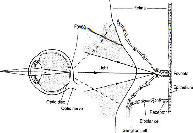
FIGURE 14-5 In most of the retina, light must first pass through overlying layers of neurons and their processes before it reaches the photoreceptors. In the center of the fovea, however, in the foveola, these neural elements are shifted aside; therefore, light has an unobstructed pathway to the photoreceptors in this region. An enlarged drawing of the foveal region is shown on the right. (From Kandel ER, Schwartz JH: Principles of neural science, ed 2, NewYork, 1985, Elsevier Science Publishing.)
because it allows light to have a less distorted path to the region of the retina associated with the highest visual acuity. The optic disc is just nasal to the fovea.