The Gastrointestinal System Has an Intrinsic Endocrine System
The GI system has an extensive number and variety of endo- crine and paracrine cells, which are distributed diffusely throughout the gut epithelium. Endocrine cells are those that produce true hormones, which by definition are molecules that travel through the blood from their site of synthesis to their site of action.
Paracrine substances, on the other hand, are molecules that are secreted by one cell and exert their effect locally, traveling by diffusion to nearby target cells. Typically, both the GI endocrine cells and the GI paracrine cells are columnar with a broad base and a narrow apex (Figure 27-7). They are positioned individually among the other mucosal cells in both the secretory and the absorptive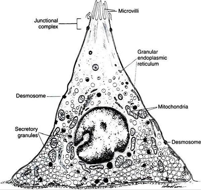
FIGURE 27-7 Gastrointestinal endocrine cell. All the Gl endocrine cells have a similar structure, but each cell produces only one type of hormone. Note the narrow apex that is exposed to the intestinal luminal contents and the broad base for storage of secretory granules. (From Johnson LR, Christensen J, Jacobsen ED, et al, editors: Physiology of the gastrointestinal tract, vol 1, ed 2, NewYork, 1987, Raven Press.)
areas of the mucosa. The narrow apex of these cells is exposed to the lumen of the gut, allowing them to “sample*’ or “taste” the luminal contents. This anatomical arrangement provides a mechanism for the cells to sense changes in gut luminal contents and to respond by releasing hormones and other regulatory substances. The base of these cells contains secretory granules, which are storage forms of hormones and paracrine substances. Endocrine cells secrete near blood vessels, whereas paracrine cells secrete into the interstitial fluid. Some paracrine cells have long basal appendages that direct their secretions to the vicinity of target cells.
It is important for students to appreciate that the secretory molecules of these cells are regulatory, not digestive. The products of these cells are secreted into either the blood or the interstitial fluid, not into the lumen of the gut. The many types of endocrine and paracrine cells in the gut epithelium are morphologically similar. However, despite their similar appearance, many distinct populations of cells produce a wide variety of hormones and other regulatory substances. Many of these substances are peptides in structure and can be referred to collectively as regulatory peptides, an inclusive term that may include molecules of neurocrine, endocrine, or paracrine origin (see previous definition of neurocrine). There are at least 28 peptides that have, or that are suspected to have, gut regulatory functions. Not all regulatory molecules in the gut are peptides, however, and several important nonpeptide paracrine and neurocrine substances exist. See Box 27-1 for a listing of peptide and nonpeptide neurocrine, paracrine, and endocrine molecules of importance in the regulation of gut function. For convenience and clarity, all these substances are frequently referred to collectively as neurohumoral regulatory molecules.
Each type of endocrine or paracrine cell has a characteristic distribution within the GI tract. For e?amp∖e, gastrin-producing cells, called G cells, are found primarily in the distal portion of the stomach, and few are found elsewhere in the gut. Cholecystokinin-producing cells are found in the small intestine, especially in the proximal region. Thus, although endocrine cells are distributed throughout the GI tract, the production of individual regulatory molecules may be confined to specific areas. This is not always the case, however, as with Somatostatin- and serotonin-producing cells, which are distributed along the entire length of the gut (Figure 27-8). Table 27-1 lists the sites of production and actions of the major GI hormones.
A subset of GI paracrine cells is known as enterochromaffin cells because of their particular histological staining characteristics. These cells are found extensively throughout the gut, although their density varies from region to region (see Figure 27-8). These mucosal cells are similar in structure to the endocrine and paracrine cells previously discussed, being exposed to the luminal contents of the gut through their apical membranes. These cells secrete a regulatory molecule known as serotonin or 5-hydroxytryptamine (5-HT). This regulatory substance is particularly important in signaling neurons that have excitatory influence on gut muscle, as well as in generating sensory signals from the gut mucosa.
The GI regulatory peptides influence various gut functions and in many cases form part of regulatory feedback loops. An
Table 27-1
Major Gastrointestinal Hormones
| Honnone | Site of | ||
| Production | Action | Release stimulus | |
| Gastrin | Distal stomach | Primary: Stimulates acid secretion from stomach glands Secondary: Stimulates gastric motility, growth of stomach epithelium | Protein in stomach; high gastric pH; vagal stimulation |
| Secretin | Duodenum | Primary: Stimulates bicarbonate secretion from pancreas Secondary: Stimulates biliary bicarbonate secretion | Acid in duodenum |
| Cholecystokinin (CCK) | Duodenum to ileum, with highest concentration in duodenum | Primary: Stimulates enzyme secretion from pancreas Secondary: Inhibits gastric emptying | Proteins and fats in small intestine |
| Gastric inhibitory | Duodenum and | Primary: | Carbohydrate and fat in small |
| polypeptide (GIP) | upper jejunum | Inhibits gastric motility and secretory activity Secondary: Stimulates insulin secretion provided sufficient glucose is present; may be most important action in many species | intestine |
| Motilin | Duodenum and jejunum | Primary: Probably regulates motility pattern of the gut in period between meals Secondary: May regulate tone of lower esophageal sphincter | Acetylcholine |
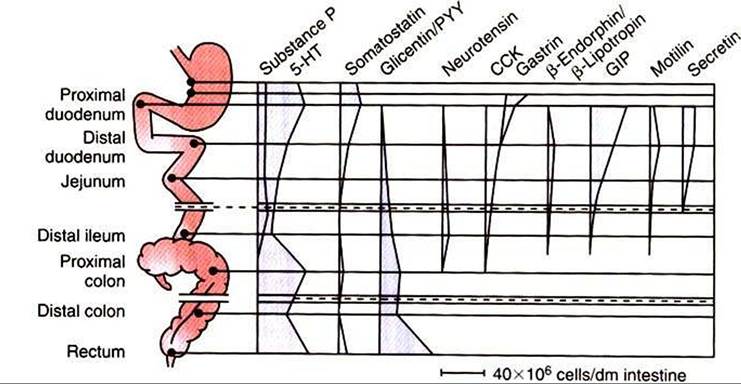
FIGURE 27-8 Distribution of gastrointestinal endocrine and paracrine cells throughout the gut, based on the hormone or paracrine substance they secrete.
(Modified from Brown DR, editor: Gastrointestinal regulatory peptides. In Handbook of experimental pharmacology, vol 106, Berlin, 1993, Springer-Verlag.)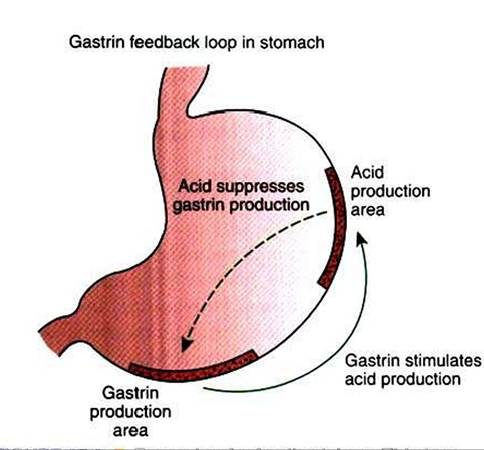
FIGURE 27-9 Example of a feedback loop.This is a negative feedback loop in that acid production, which is stimulated by gastrin, suppresses gastrin secretion.
example is the feedback loop that involves gastrin and gastric acid. After a meal, gastrin-producing cells, which are located in the antrum of the stomach, secrete gastrin. Gastrin then acts as a hormone traveling through the blood to stimulate gastric acid production from specialized cells in the body of the stomach. The acid is secreted into the lumen of the stomach, lowering the pH of stomach contents. The feedback loop is completed as acidified stomach contents come into contact with the apical portion of the G cells. The reduced pH of the stomach contents signals the cells to stop gastrin secretion, removing a major stimulus for the production of gastric acid and thus stabilizing stomach pH. The actions of the G cells therefore closely regulate the pH of the stomach (Figure 27-9). Throughout this discussion of the GI system, you will see that this general pattern of endocrine or paracrine stimulus and feedback inhibition is inherent in the function of most GI regulatory peptides and other molecules. Other specific examples of Gl neurohumoral regulatory effects are discussed in subsequent chapters, in the context of the actions they regulate.