The Gut Receives Extrinsic Innervation from the Autonomic Nervous System
The parasympathetic and sympathetic nervous systems form the link between the central nervous system (CNS) and the ENS. Most of the GI tract receives parasympathetic innervation by way of the vagus nerve, except the terminal portions of the colon, which receive parasympathetic innervation from the sacral cord through the pelvic nerve (Figure 27-5).
By classic description, the parasympathetic nervous system is composed OfpregangIionic and postganglionic fibers (see Chapter 13). However, this division of parasympathetic fiber types is not clearly defined for the gut, because extrinsic preganglionic fibers of the parasympathetic system become integrated with fibers of the ENS. Parasympathetic, preganglionic fibers reach the gut and synapse on cell bodies of the ENS, and thus the enteric ganglia of the gut serve as peripheral autonomic ganglia of the parasympathetic system. However, the ENS is much more than postganglionic parasympathetic neurons. Unlike typical postganglionic parasympathetic neurons, the ENS neurons receive input from sources other than preganglionic parasympathetic fibers. These include afferent neurons and interneurons of the ENS, as well as humoral (chemical) influences from other cells in the gut (see next section). Thus the classic description of the organization of the parasympathetic nervous system does not strictly apply to the gut (Figure 27-6).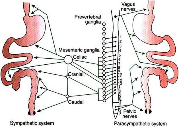
FIGURE 27-5 Distribution of autonomic nerve fibers to the gut.The spinal cord is represented in the center, with the sympathetic system extended to the left and the parasympathetic system to the right.
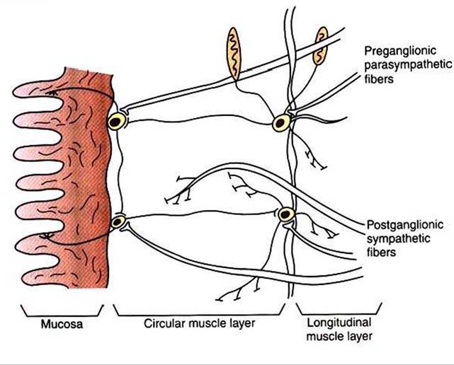
FIGURE 27-6 Interface between the autonomic and enteric nervous systems. Note that parasympathetic fibers reaching neurons of the enteric system are preganglionic, whereas sympathetic fibers are postganglionic.
In contrast to the extrinsic parasympathetic fibers, extrinsic sympathetic fibers that enter the gut are primarily postganglionic. Postganglionic sympathetic fibers arise from cells in the prevertebral ganglia (see Chapter 13) and follow the splanchnic nerves and vascular arteries into the gut wall (see Figure 27-5). Some sympathetic fibers synapse on neurons of the ENS, whereas others exert a direct effect on GI muscles and glands.
Neurons of the sympathetic and parasympathetic nervous system influence GI function through the release of neuro- crines. In general, parasympathetic neurocrines are stimulatory (i.e., they increase gut blood flow, motility, and glandular secretions), whereas those of the sympathetic system are inhibitory.