The heart is almost completely invested by and fits snugly in the pericardium (Fig. 7.5).
The pericardium makes a serous sac, but its very tight fit around the heart reduces the lumen to a mere capillary space that contains a small amount of serous fluid to facilitate the easy movement of the heart wall in the pericardial sac (Fig.
7.5/4). The visceral and parietal layers of the pericardium continue into each other at a complicated reflection that runs over the atria and the roots of the great vessels. The visceral layer is so closely adherent to the epicardium that it may be described as an epicardial component. The parietal layer has a thick external fibrous covering (Fig. 7.5/6) that blends with the adventitia of the great vessels dorsally and forms a ligament at the ventral apex of the sac to attach to the sternum (sternopericardial ligament; Fig. 7.5/8) or to the diaphragm (phrenicopericardial ligament). These attachments place a severe restraint on the mobility of the heart, although slight movement does occur with each respiratory excursion.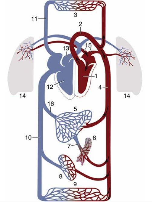
FIG. 7.4 Schematic drawing of the systemic and pulmonary circulation. 1, Left ventricle; 2, aorta; 3, capillary bed of head, neck, and forelimb; 4, abdominal aorta; 5, liver; 6, capillary bed of intestines; 7, portal vein; 8, capillary bed of kidneys; 9, capillary bed of caudal part of the body; 10, caudal vena cava; 11, cranial vena cava; 12, right ventricle; 13, pulmonary trunk; l4, capillary bed of lungs; 15, pulmonary vein; 16, hepatic veins.
Although the pericardium distorts to accommodate the changing form of the heart during the cardiac cycle, its fibrous component prevents any significant distention in the short term. It may stretch over longer periods if the heart becomes enlarged by exercise or disease or if an effusion or accumulation of inflammatory fluid occurs within the pericardial cavity.
The heart (within the pericardium)* is included within the mediastinum, the partition that separates the right and left pleural cavities (see Fig. 4.20A). It is conical and is placed asymmetrically within the thorax, and the larger part (about 60%) lies to the left of the median plane (see Figs. 13.13B and 20.8). The base is dorsal and reaches approximately to the horizontal (dorsal) plane that bisects the first rib; in some species (e.g., the dog) it is tilted in varying degrees to face craniodorsally. The apex is placed close to the sternum, opposite the sixth costal cartilage. The long axis that joins the center of the base to the apex thus slopes caudoventrally, with some deviation to the left imposed by the skewed orientation (Fig. 7.6). The projection of the heart on the chest wall extends between the third and sixth ribs (or thereabouts). Thus, much of the heart is covered by the forelimb, which makes clinical examination a challenge, especially in larger animals (See Chapters 20 and 27).
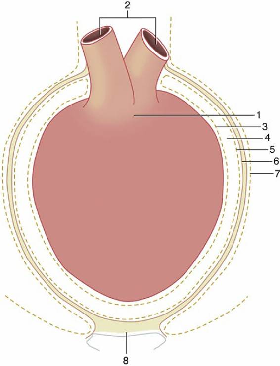
FIG. 7.5 Schematic illustration of the pericardium. 1, Heart; 2, great vessels; 3, visceral pericardium (epicardium); 4, pericardial cavity (exaggerated in size); 5, parietal pericardium; 6, connective tissue layer of the parietal pericardium; 7, mediastinal pleura; 8, sternopericardial ligament.
The heart displays some lateral compression to conform to the shape of the thorax of most quadrupeds. This shape of the heart better defines right and left surfaces (which are also crossed by the corresponding phrenic nerves) make a better fit with the corresponding lungs. The cardiac notch in the ventral border of each lung allows the heart a restricted contact with the chest wall, which is normally greater on the left side because of the asymmetrical position (see Fig. 13.5). The cranial aspect is extensively related to the thymus (in the young animal), but the caudal surface faces toward the diaphragm and may be indirectly related through it to cranial abdominal organs a point of importance in certain species such as cattle (see Chapter 28).
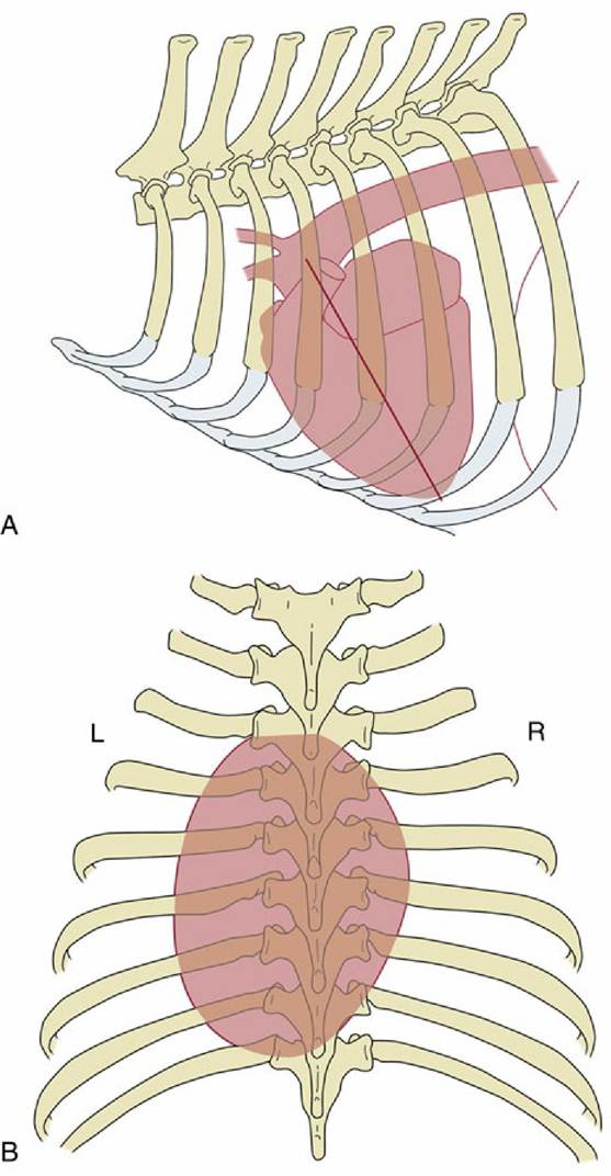
FIG. 7.6 Schematic drawings to show the position of the canine heart, based on radiographs. (A) Left lateral view; the caudoventrally sloping long axis (redline) of the heart is indicated. (B) Dorsoventral view showing the asymmetrical position of the heart. L, left; R, right.