The heart (cor) is the central organ that pumps blood continuously through the blood vessels by rhythmic contraction.
The adult heart has right atrium, left atrium, right ventricle, and left ventricle (Fig. 7.3). The two atria and the two ventricles are separated by an internal septum, but the atrium and ventricle of each side communicate through a large opening.
The heart consists of two pumps that are combined within a single organ. The deoxygenated (venous) blood enters the right atrium and is ejected to the lungs via the pulmonary trunk. The left pump receives the oxygenated blood from the lungs via the pulmonary veins and ejects it into the aorta for distribution to the body (Fig. 7.4).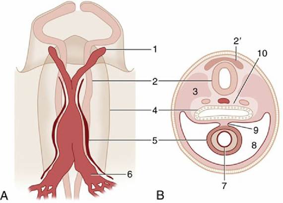
FIG. 7.1 (A) Ventral view of the cranial part of a 15-day-old pig embryo after fusion of the endocardial tube. (B) Transverse section of a seven- to eight-somite embryo taken at the level of 5. 1, First aortic arch; 2, neural tube; 2', neural crest; 3, somite; 4, foregut; 5, epimyocardial wall of the fused endocardial tubes; 6, vitelline vein; 7, endocardial tube; 8, pericardial cavity; 9, dorsal mesocardium; 10, notochord and dorsal aortae.
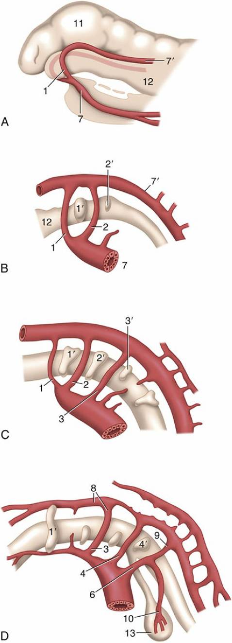
FIG. 7.2 (A) to (D) Left lateral views of the aortic arches and their transformation. (A) Dorsal and ventral aortae are connected by the first aortic arches. (B) First and second aortic arches are present. (C) The first arch begins to disappear, the third is complete, and the fourth and sixth develop. (D) The third arch
and the cranial part of the dorsal aorta are now transformed into the internal carotid artery, and the sixth gives rise to the pulmonary trunk and ductus arteriosus. 1-4 and 6, Aortic arches; 1'-4', pharyngeal pouches; 7 and 7', ventral and dorsal aortae; 8, internal carotid artery; 9, ductus arteriosus; 1θ, left pulmonary artery; 11, brain vesicle; 12, foregut; 13, lung bud.
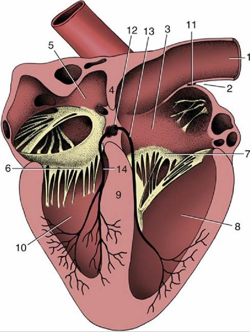
FIG. 7.3 Section of the heart exposing the four chambers. 1, Cranial vena cava; 2, terminal sulcus; 3, right atrium; 4, interatrial septum; 5, left atrium; 6, left atrioventricular valve; 7, right atrioventricular valve; 8, right ventricle; 9, interventricular septum; 10, left ventricle; 11, sinoatrial node; 12, atrioventricular node; 13 and 14, right and left limbs of atrioventricular bundle.
The size of the heart varies considerably among species and also among individuals but generally forms 0.75% of the body weight. The heart is typically and relatively larger in smaller species and animals of smaller size but larger in athletic animals such as the Thoroughbred horse and Greyhound. The heart becomes larger (hypertrophied) with exercise. However, the construction, the form, and the general position of the heart are similar among mammals. The differences in topography are mentioned in later chapters because of their significance in clinical examination.