THE HEART (See also pp. 228-234.)
The canine heart is ovoid. Its long axis forms an angle of about 45° with the sternum; the base thus faces cra- niodorsally, and the blunt apex lies near the junction of the sternum and the diaphragm, a little to the left of the midline (Figure13-17, A-B).
The angle between the axis of the heart and the sternum and the space between the apex and the diaphragm vary both more considerably than many accounts suggest. The angle is greater and the shape of the heart more conical in deep-chested breeds. Because the position of the heart is biased, a thinner layer of lung tissue intervenes between the heart and the left thoracic wall, resulting in the heart sounds being more pronounced on the left side (see Figure 13-10, Figure 13-17, A-B, and Figure 13-21, A-B).The heart contributes about 0.7% of the body weight on average, but its weight, both absolute and relative, varies considerably. Dogs trained for hunting or racing have hearts two or three times heavier than those of fat and less athletic individuals of comparable size.
The left surface presents the auricles embracing the pulmonary trunk, and below the coronary groove the ventricles are divided by the paraconal interventricular groove (see Figure 13-14). The right surface presents the atria and the subsinuosal interventricular groove. Neither surface faces quite as its name suggests; the left surface is rotated a little more toward the sternum, and the right one is a little more toward the vertebrae. When one reads counterclockwise from the base, the periphery of the heart shadow in a left lateral radiograph presents the right auricle, the right ventricle, the left ventricle, and the left atrium (Figure 13-17/1-4); in a ventrodorsal radiograph the sequence is right atrium, right ventricle, left ventricle, and pulmonary trunk (Figure 13-17/2,3,5,6). The apex is formed only by the wall of the left ventricle.
It is clearly important to know the relationships of the parts of the heart to external landmarks. The heart extends from the third rib to the sixth intercostal space, and the latter limit roughly coincides with the most
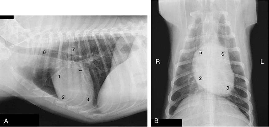
Figure 13-17 Lateral (A) and ventrodorsal (B) views of the position of the canine heart. 1, Right auricle; 2, right ventricle; 3, left ventricle; 4, left atrium; 5, right atrium; 6, pulmonary trunk; 7, aorta; 8, trachea.
cranial extent of the diaphragm (Figure 13-17, A). The projection of the base intersects the middle of the fourth rib; the most dorsal part of the heart reaches approximately to the line connecting the acromion with the ventral end of the last rib. The apex lies just to the left of the second last sternebra. In the standing dog the apex beat is palpable on both sides, low in the fifth or sixth intercostal space. The main contractions are said to be strongest in the lower third of the fourth or fifth space and to be a little more pronounced on the left. The ductus arteriosus or its replacement, the ligamentum arteriosum (p. 255), is located where the pulmonary trunk is intersected by the left vagus, opposite the fourth rib (see Figure 13-14). These details are relevant to the diagnosis and surgical treatment of persistent ductus arteriosus, the most common congenital anomaly of the canine cardiovascular system. Among other signs, a persistent ductus produces a characteristic “machine” murmur. The condition can be treated by ligation and section of the duct. It may be reached by a left lateral thoracotomy with the use of the fourth intercostal space. The same approach provides access to the right ventricle, left auricle, pulmonary trunk, and descending aorta. (The fourth space on the right side may be used to gain access to the main part of the right ventricle, both atria, the ascending aorta, and both the caval and the azygous veins.)
The heart is more easily auscultated than in the larger species because it is less covered by the forelimbs and a stethoscope can be introduced deeply into the axilla.
The puncta maxima for optimal perception of the valve sounds may be summarized: the left atrioventricular valve—low (at the costochondral junction) in the left fifth intercostal space; the pulmonary valve—low in the left third space; the aortic valve—high (just below the horizontal plane of the shoulder joint) in the left fourth space; the right atrioventricular valve—high (just a little lower than the location of the aorta at the left side) in the fourth space on the right side (Figure 13-5, A-D). These findings correspond surprisingly closely with those determined at postmortem examinations of dogs diagnosed in life as having valvular lesions, despite the distorting influence of tissues on the conduction of sound.There are no significant structural peculiarities of the canine heart, although it may be noted that the right atrioventricular valve possesses only two major cusps in many (perhaps most) dogs. No clinical significance attaches to the variation.
In North America many dogs are infested with large heartworms (Dirofilaria immitis), which occupy the pulmonary trunk and, in severe cases, the right ventricle, atrium, and caudal vena cava.
The heart of the cat extends from the third (or fourth) to the sixth (or seventh) rib. Little is covered by the forelimb in the standing animal because the triceps reaches no farther than the fourth rib. The long axis of the heart forms a more acute angle with the sternum, which results in a greater area of sternal contact than in most dogs. The contractions are strongest near the ventral ends of the fourth to sixth ribs on the left and the fifth rib on the right (Figure 13-18). The corresponding puncta maxima are as follows: the left atrioventricular valve—in the fifth and sixth intercostal space, level with the shoulder joint; the pulmonary and aortic valves—low in the left second and third intercostal space; and the right atrioventricular valve—level
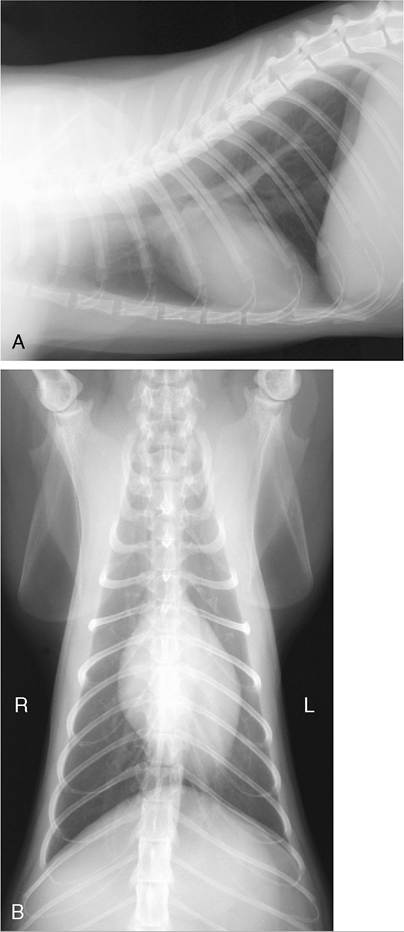
Figure 13-18 Lateral (A) and ventrodorsal (B) radiographic views showing the position of the feline heart. The ventral ends of ribs 5, 6, and 7 lie on the heart shadow in A.
with the shoulder joint in the fourth and fifth intercostal space. Puncture is difficult because the organ is so small; a needle inserted on either side of the right fifth costochondral junction should enter a ventricle.