THE MEDIASTINUM (See also pp. 158-160.)
The fibrous tissue associated with the thoracic organs and between the pleural sacs (fascia endothoracica) is
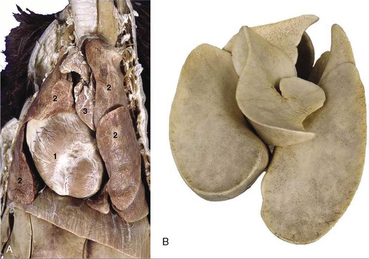
Figure 13-11 A, Thoracic viscera of the dog.
1, Heart; 2, pulmonary lobes; 3, thymus. In B (inflated specimen) the deep fissure between the lobes of the lung are clearly visible.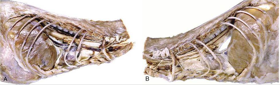
Figure 13-12 A, Mediastinum of a cat, right view. In the middle part the heart is the main component. The cranial and caudal mediastinum is thin and, in some places, fragile. B, Mediastinum, left view. A large opening in the caudal part, probably caused by dissection, indicates the fragility of the structure.
so thin that the mediastinum is reduced in several places to a very delicate and transparent membrane (Figure 13-12, B) consisting only of apposed right and left pleural sheets. It ruptures easily, and although the two pleural sacs may be regarded as normally independent, most dogs in which pneumothorax has been induced unilaterally show bilateral pneumothorax in radiographs.
The cranial mediastinum is wide dorsally where it contains the trachea and esophagus lying side by side as they pass through the thoracic inlet; below these the cranial vena cava and brachiocephalic trunk, with their tributaries and branches, are embedded in generous quantities of fat. Ventrally the cranial mediastinum contains lymph nodes, the internal thoracic vessels, fat, and, in the young animal, the thymus. This part narrows with the regression of the thymus, providing more space for the apices of the lungs.
The dorsal part of the middle mediastinum is slightly narrower than the heart (Figure 13-13); it
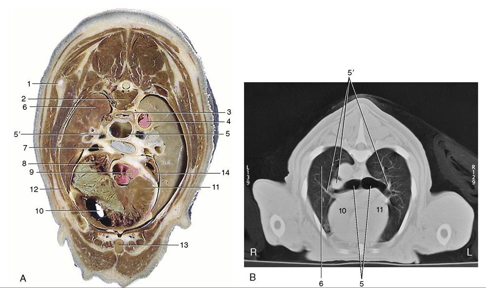
Figure 13-13 A, Transverse section of the canine trunk at the level of the sixth thoracic vertebra.
B, Corresponding computed tomographic image at a slightly more caudal level. 1, Caudal angle of scapula; 2, sixth thoracic vertebra; 3, esophagus; 4, aorta; 5, tracheal bifurcation; 5', large blood vessels accompanying principal bronchi are likely right and left pulmonary aa.; 6, right lung; 7, tracheobronchial lymph nodes and pulmonary a.; 8, right atrium; 9, origin of aorta; 10, right ventricle; 11, interventricular septum; 12, fifth rib; 13, sternum; 14, left auricle.contains the termination of the trachea, the esophagus, the aortic arch, the structures comprising the roots of the lungs, and lymph nodes. Its right surface is flat, but the aorta (Figure 13-13Z√) bulges laterally on the left, indenting the left lung. The middle part at this level contains the heart (within the pericardium), while the ventral part, between the pericardium and sternum, is folded, resembling the greater omentum, and empty but for the phrenicopericardiac ligament, which attaches the pericardium to the sternum and diaphragm more loosely than the tether provided by the corresponding sternopericardiac ligament of the larger species.
The triangular dorsal part of the caudal mediastinum contains the aorta and the right azygous vein and, more ventrally, the esophagus (Figure 13-12, A, through Figure 13-16). The delicate ventral part runs between the pericardium and the diaphragm, which it approaches along a line that is displaced so far to the left that it reaches the thoracic wall near the ninth costochondral junction. There is the usual recess between the mediastinum and the fold enclosing the caudal vena cava that is occupied by the accessory lobe of the right lung.
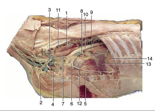
Figure 13-14 Left lateral view of the canine thoracic cavity; the lung and much of the pericardium have been removed. 1, Longus colli; 2, left subclavian artery; 3, internal thoracic vessels; 4, thymus; 5, vessels in paraconal interventricular groove; 6, pulmonary trunk; 7, esophagus; 8, pulmonary veins entering left atrium; 9, left principal bronchus and dorsal and ventral vagal trunks; 10, aorta; 11, sympathetic trunk; 12, phrenic nerve; 13, caudal mediastinum; 14, diaphragm.
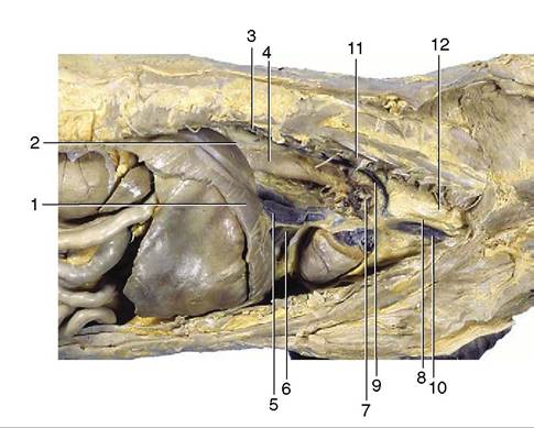
Figure 13-15 Right lateral view of the canine thoracic cavity; the lung and much of the pericardium have been removed.
1, Diaphragm; 2, infracardiac bursa; 3, sympathetic trunk; 4, esophagus; 5, caudal vena cava; 6, plica venae cavae; 7, root of lung and phrenic nerve; 8, right vagus; 9, right azygous vein; 10, cranial vena cava; 11, longus colli; 12, trachea.A diverticulum of peritoneum, the infracardiac bursa, intrudes through the esophageal hiatus of the diaphragm to lie against the right face of the esophagus, extending from the diaphragm to the root of the lung. It is the occasional recipient of a herniated part of an abdominal organ, either as a congenital anomaly or as the result of trauma.
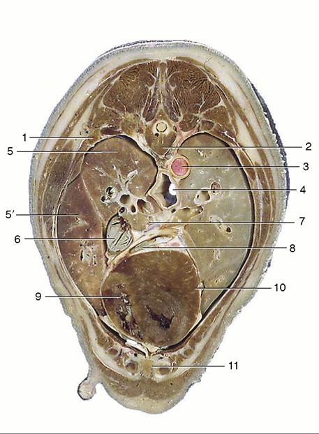
Figure 13-16 Transverse section of the canine trunk at the level of the seventh thoracic vertebra. 1, Sixth rib; 2, seventh thoracic vertebra; 3, aorta; 4, esophagus; 5, cranial lobe; 5', middle lobe of right lung; 6, caudal vena cava; 7, pulmonary veins passing to left atrium; 8, great cardiac vein; 9, right ventricle; 10, left ventricle; 11, sternum.