The Hindbrain
The hindbrain (rhombencephalon) comprises the medulla oblongata, pons, and cerebellum. These parts differentiate from the caudal brain vesicle shortly after closure of the neural tube.
Thinning of the roof plate in this region weakens the structure and causes the vesicle to flatten as the pontine flexure develops. The flattening splays the lateral wall of the neural tube outward so that the luminal surfaces come to face dorsomedially; the alar plates are now lying lateral to the basal plates (Fig 8.24). The part of the hindbrain caudal to the pontine flexure (the myelencephalon) becomes the medulla oblongata of adult anatomy. The rostral part is the metencephalon, which becomes the pons and cerebellum in the adult. The parts of the roof plate caudal and rostral to the cerebellum remain thin and form the rostral and caudal medullary vela (velum, singular) that form the roof of the lumen, known as the fourth ventricle in the adult (see Fig. 8.24).The Medulla Oblongata and Pons
The medulla oblongata and pons together form the caudal regions of the brainstem, and internally there is no distinct division between them. Externally, the pons corresponds in extent to the large transverse group of axons that encloses its ventral and lateral aspects and continues into the cerebellum as the middle cerebellar peduncles (see Fig. 8.22/9).
Although the medulla oblongata continues the spinal cord caudally, it widens toward its rostral end as the result of the developmental flattening of the myelencephalon. The medulla oblongata's ventral surface is marked by a median fissure continuous with that of the cord and flanked by longitudinal ridges, the pyramids (Fig. 8.19/17). Many of the constituent fibers of the pyramids decussate (cross to the opposite side) at the transition of spinal cord and medulla, forming interlacing bundles within the fissure.
More cranially, a lesser transverse ridge, the trapezoid body, crosses the ventral surface of the medulla oblongata directly caudal to the transverse fibers of the pons. The other noteworthy features on the ventral surface of the pons and medulla are the superficial origins of many of the cranial nerves. The trigeminal nerve (V) appears at the lateral aspect of the transverse pontine fibers; the abducent nerve (VI) emerges caudal to this and more medially, through the trapezoid body lateral to the pyramid; the facial (VII) and vestibulocochlear (VIII) nerves appear to continue the trapezoid body laterally; the glossopharyngeal (IX), vagus (X), and accessory (XI) nerves arise from the lateral aspect of the medulla oblongata in close succession; and the hypoglossal nerve (XII) takes a more ventral origin in line with that of the abducent nerve and the ventral roots of the spinal nerves (Figs. 8.19 and 8.20).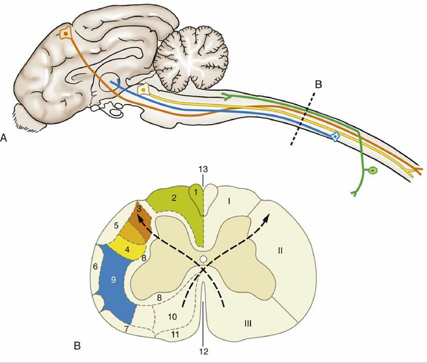
FIG. 8.18 (A) Principal ascending and descending tracts of the spinal cord, showing the locations of the cell bodies and axon pathways. (B) Hypothetical transverse section at the level indicated shows the the relative location of principal tracts. The curved dotted arrows indicate the crossing of the pyramidal tracts, which occurs at the juncture of the brain and spinal cord. (The drawing has been simplified for the sake of clarity.) I, Dorsal funiculus; II, lateral funiculus; III, ventral funiculus; 1, fasciculus gracilis; 2, fasciculus cuneatus; 3, lateral corticospinal tract; 4, rubrospinal tract; 5, dorsal spinocerebellar tract; 6, ventral spinocerebellar tract; 7, spino-olivary and olivospinal tracts; 8, propriospinal system (fasciculi proprii); 9, spinothalamic tract; 10, ventral corticospinal tract; 11, vestibulospinal tract; 12, ventral median fissure; 13, dorsal median sulcus.
It is helpful to study a median section of the brain (Fig. 8.21) before examining the dorsal aspect of the medulla oblongata and pons.
This section shows that the fourth ventricle is located close to the dorsal surface of the brainstem. The ventricle is covered by a tented roof formed in part by the ventral surface of the cerebellum and in part by the rostral and caudal medullary vela (Fig. 8.21/15 and 15'), which extend from the cerebellum to the midbrain and from the cerebellum to the closed caudal part of the medulla oblongata, respectively. Exposure of the dorsal surface of the medulla and pons requires the removal of the cerebellum by transection of its peduncles attaching it to the pons, an operation that almost inevitably destroys the fragile vela (Fig. 8.22).The fourth ventricle is diamond shaped from a dorsal view (see Fig. 8.22) and is aptly named the rhomboidal fossa; its widest part is at the pontine-medullary junction. The margins of the fossa are provided by the three pairs of cerebellar peduncles. The floor is irregular and is marked by a median sulcus on the midline and paired lateral (limiting) sulci. The most rostral part of the rostral velum, a part that occasionally survives removal of the cerebellum, contains the superficial origins of the trochlear nerves (IV), the only nerves to emerge from the dorsal aspect of the brain.
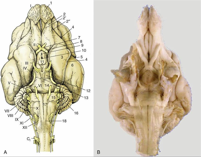
FIG. 8.19 (A) Ventral view of the canine brain. 1, Olfactory bulb; 2, olfactory tract; 2', medial olfactory tract; 2", lateral olfactory tract; 3, piriform lobe; 4, rhinal sulcus; 5, sylvian sulcus; 6, ectosylvian gyrus; 7, optic chiasm; 8, optic tract; 9, tuber cinereum; 10, infundibulum (the hypophysis has been detached and the third ventricle is opened); 11, mammillary body; 12, crus cerebri; 13, interpeduncular fossa; 14, pons; 15, trapezoid body; 16, cerebellar hemisphere; 17, pyramidal tract; 18, crossing of pyramidal tracts. I-XII designate the appropriate cranial nerves; C1, Cervical nerve 1. (B) The real specimen of the dog.
On either side of the caudal part of the fourth ventricle, the dorsal surface of the medulla oblongata is represented by inconspicuous eminences, the gracile and cuneate nuclei (Fig.
8.23/5 and 7), which are the cranial terminations of the similarly named gracile and cuneate fasciculi of the dorsal funiculus of the spinal cord. These nuclei transmit somatosensory information from the body and limbs, as discussed later in the chapter.The principal features of the internal anatomy of the medulla oblongata and pons are as follows: the nuclei of the cranial nerves, the olivary and pontine nuclei, the reticular formation, and certain ascending and descending fiber tracts that connect the spinal cord with higher regions of the brain. These structures are described here but without excessive attention to establishing their topographic relationships.
The Nuclei of the Cranial Nerves
The nuclei of the cranial nerves represent the continuation of the four functional components, somatic afferent, visceral afferent, visceral efferent, and somatic efferent, that compose the gray matter of the spinal cord (see Fig. 8.12). These components are supplemented by two additional components, special somatic afferent and special visceral afferent, that are carried by cranial nerves in connection with the innervation of receptors for special senses, which have no counterparts in the trunk or limbs (i.e., hearing, balance, taste).
Recall that in the spinal cord, the regions receiving somatic and visceral afferent input are located together in the dorsal gray column whereas the somatic and visceral motor neurons are located in the ventral column. In the medulla, the widening of the roof of the fourth ventricle and flattening of the brainstem cause these components to be arranged medial to lateral (see Fig. 8.12). These components now exhibit a lateromedial rather than dorsoventral sequence, with a lateral somatic afferent column and a medial somatic efferent column. Certain of the columns also fragment into discrete parts (nuclei), and at some levels the relationships are further adjusted to allow the intrusion of the additional components. The consequences are that those cranial nerves that contain more than one functional component arise from more than one nucleus and that certain nuclei give rise to similar components of more than one nerve.
The general arrangement of the six components is illustrated in Fig. 8.25 in a schematic fashion.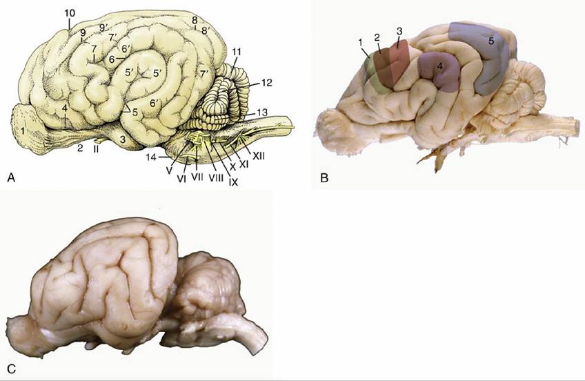
FIG. 8.20 (A) Lateral view of the canine brain. 1, Olfactory bulb; 2, olfactory tract; 3, piriform lobe; 4, rhinal sulcus; 5, sylvian sulcus; 5', sylvian gyrus; 6, ectosylvian sulcus; 6', ectosylvian gyrus; 7, suprasylvian sulcus; 7', suprasylvian gyrus; 8, ectomarginal sulcus; 8', ectomarginal gyrus; 9, coronal sulcus; 9', coronal gyrus; 10, cruciate sulcus; 11, cerebellar vermis; 12, cerebellar hemisphere; 13, paraflocculus; 14, pons; II and V-XII designate the appropriate cranial nerves. (B) Lateral view of the canine brain. 1, motor cortex; 2, overlap of motor and somatosensory cortex; 3, somatosensory cortex; 4, auditory cortex; 5, visual cortex (C) Lateral view of the feline brain.
The somatic efferent column serves muscles that have originated from somites and branchiomeres of the head. Its medial part is fragmented into a long hypoglossal nucleus and a smaller abducent nucleus within the floor of the fourth ventricle (and trochlear and oculomotor nuclei within the tegmentum of the midbrain). The fibers from the oculomotor, abducent, and hypoglossal nuclei take the expected courses to emerge on the ventral aspect of the brain, close to the midline and in line with one another and the ventral roots of the spinal nerves (see Fig. 8.19). Those that compose the trochlear nerve emerge from the dorsal aspect of the brain after decussation within the rostral medullary velum (Fig. 8.22/IV); this is an aberrant course for a cranial nerve, for which there is no satisfactory explanation.
The lateral (branchiomeric) portion of the somatic efferent column (see Fig. 8.25) supplies the striated masticatory, facial, laryngeal, and pharyngeal muscles through the trigeminal, facial, glossopharyngeal, vagus, and accessory nerves. This portion is divided into the motor nuclei of the trigeminal and facial nerves (Fig.
8.25/16 and 17) and the nucleus ambiguus (Fig. 8.25/14) shared by the glossopharyngeal and vagus nerves. The fibers emerge from the ventrolateral surface of the brainstem but do not always take the most direct internal course to do so.The visceral efferent column supplies the autonomic (parasympathetic) motor component of certain cranial nerves. The lateral of the efferent columns (Fig. 8.24/4), it is divided into the parasympathetic nucleus of the vagus nerve (Fig. 8.25/13), the caudal salivatory nucleus of the glossopharyngeal nerve, and the rostral salivatory nucleus of the facial nerve (Fig. 8.25/15) (and, in the midbrain, the parasympathetic nucleus of the oculomotor nerve [Fig. 8.25/18]). The vagal parasympathetic fibers are distributed to the cervical, thoracic, and abdominal (but not pelvic) viscera, whereas parasympathetic fibers in the glossopharyngeal and facial nerves are distributed to glands of the head. Further rostrally, parasympathetic fibers within the oculomotor nerve innervate the smooth intrinsic muscles of the eyeball.
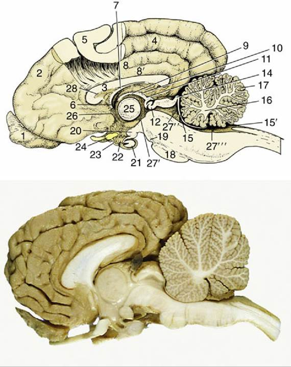
FIG. 8.21 Median section of the canine brain. Part of the medial wall of the hemisphere has been removed. 1, Olfactory bulb; 2, hemisphere; 3, corpus callosum; 4, splenial sulcus; 5, cerebral cortex; 6, interventricular foramen; 7, fornix; 8, cingulate gyrus; 8', supracallosal gyrus; 9, thalamus; 10, epithalamus; 11, epiphysis; 12, posterior commissure; 13 and 14, commissures of rostral and caudal colliculi; 15, rostral medullary velum; 15', caudal medullary velum; 16, corpus medullare; 17, cerebellar cortex; 18, pons; 19, crus cerebri; 20, mammillary body; 21, hypophysis; 22, infundibulum; 23, tuber cinereum; 24, optic chiasm; 25, interthalamic adhesion; 26, anterior commissure; 27', third ventricle; 27", mesencephalic aqueduct; 27"', fourth ventricle; 28, septum telencephali (pellucidum).
The visceral afferent column (Fig. 8.24/2 and 3) is in fact double and is made up of neurons receiving either visceral or special visceral afferent inputs. It forms a single very long nucleus (nucleus of the solitary tract [Fig. 8.25/10]) that is subdivided in relation to the associated facial, glossopharyngeal, and vagus nerves. Many neurons receive visceral input from the caudal part of the mouth and the cervical, thoracic, and abdominal viscera; the special component, which is concerned with taste, is distributed among the facial, glossopharyngeal, and vagus nerves.
The somatic afferent column (Fig. 8.24/1) extends from the cervical part of the spinal cord through the medulla and pons into the mesencephalon. It is broken into several nuclei. The rostralmost extent, the mesencephalic nucleus of the trigeminal nerve (Fig. 8.25/7), is concerned with proprioception; it presents a unique feature, the inclusion of the primary afferent neuron cell bodies within the central nervous system (the one exception to an otherwise inviolable rule that the cell bodies of primary afferent neurons are located within peripheral ganglia). The two exteroceptive nuclei (Fig. 8.25/7) are the principal sensory nucleus of the trigeminal nerve within the pons and the nucleus of the descending (spinal) tract of the trigeminal nerve, which extends from the level of the pons into the cervical part of the spinal cord.
The special somatic afferent column is associated with the optic and vestibulocochlear nerves and therefore with the special somatic senses of vision (II), balance (vestibular division of VIII), and hearing (cochlear division of VIII) (Fig. 8.25/6, 8, and 9). The afferent pathways of these important senses are considered elsewhere; our present purpose is to locate the relevant nuclei within the brainstem. The four closely related vestibular nuclei are spread through part of the medulla oblongata and pons, medial to the caudal cerebellar peduncle. The two (dorsal and ventral) cochlear nuclei are located within the most rostral part of the medulla oblongata close to the entry of the eighth nerve.
The fiber composition of the nerves is summarized conveniently in Table 8.2.
Other Internal Features
The olivary nuclear complex occupies a position in the caudal part of the medulla oblongata, dorsolateral to the pyramidal tract, where it sometimes raises a gentle surface swelling (Fig. 8.26/10). It is composed of several parts and varies considerably in form among species, generally taking the form of a nuclear lamina folded onto itself to form a bag. It has an important function in the regulation of motor feedback (pp. 288-289). Several other nuclei within the pons (Fig. 8.27) are also concerned with motor control.
The reticular formation is a diffuse system of nuclei and fiber tracts (Figs. 8.26/8 and 8.28/13) that extends from the spinal cord to the forebrain and occupies a large part of the core of the medulla oblongata and pons. It is discussed on p. 284.
The principal fiber tracts that pass through the pons and medulla are summarized here but are discussed later in this chapter along with their functional morphology. The large descending corticospinal tract that forms the pyramid on the ventral surface of the medulla (Fig. 8.26/11) and the ascending tract known as the medial lemniscus (Fig. 8.28/9) are prominent in transverse sections. The medial lemniscus consists of axons arising from neurons in the gracile and cuneate nuclei, axons that initially travel ventrally from the nuclei (as the deep [internal] arcuate fibers), then cross the midline in the ventral part of the caudal medulla before turning rostrally as the prominent medial lemniscal bundle. Also traversing the pons and medulla are axons of the trigeminothalamic and cervicothalamic tracts, which arise from the principal sensory nucleus of the trigeminal nerve and the lateral cervical nucleus in the spinal cord, respectively. The three cerebellar peduncles are also prominent here, the composition, origin, and destination of which are described later.
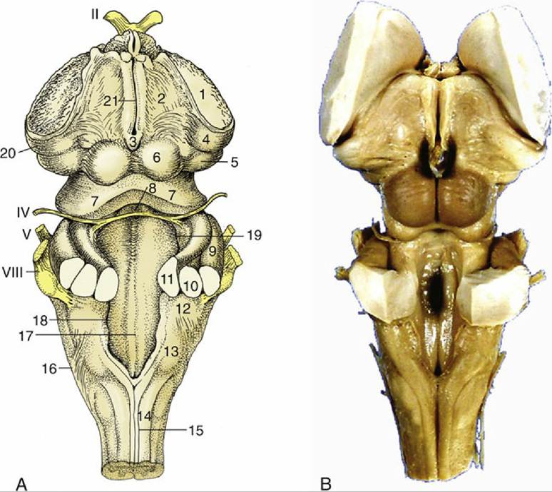
FIG. 8.22 (A) Dorsal view of the canine brainstem with the cerebellum removed and the fourth ventricle opened. 1, Cut fibers of internal capsule; 2, dorsal part of thalamus; 3, epiphysis; 4, lateral geniculate body; 5, medial geniculate body; 6, rostral colliculus; 7, caudal colliculus; 8, decussating fibers of trochlear nerves in the rostral velum; 9, middle cerebellar peduncle; 10, caudal cerebellar peduncle; 11, rostral cerebellar peduncle; 12, dorsal cochlear nucleus; 13, cuneate tubercle; 14, fasciculus cuneatus; 15, fasciculus gracilis; 16, superficial arcuate fibers; 17, median sulcus; 18, medial eminence; 19, sulcus limitans; 20, optic tract; 21, margin of roof of third ventricle; IV, V, and VIII designate the appropriate cranial nerves. (B) Dorsal view of equine brainstem.
The Cerebellum
The cerebellum is a roughly globular, multiply and deeply fissured mass that is located above the pons and medulla oblongata and is connected to the brainstem by three peduncles on each side (Fig. 8.22/9-11). It is separated from the cerebral hemispheres cranially by the transverse fissure occupied by the membranous tentorium cerebelli (p. 295) when the brain is in situ in the skull.
The cerebellum consists of two large lateral hemispheres and a narrower median region named the vermis owing to its supposed resemblance to an earthworm. Deep to both of these regions, a small flocculonodular lobe is separated by deep fissures from the larger mass (see Fig. 8.20). Smaller fissures divide each region into lobules and these into yet smaller units known as folia. The lobules are individually named, but neither their names nor their exact forms are important.
The arrangement of the gray matter and white matter sharply contrasts that found in the spinal cord and medulla oblongata. In the cerebellum the bulk of the gray matter is arranged as an external cortex that encloses the white matter, or "medulla" (see Fig. 8.21). The cortex is highly folded to accommodate the large amount of gray matter required for neural processing. The medulla, consisting of myelinated axon tracts arising from the peduncle and radiating through the various lobes, lobules, and folia, form a branching structure with some resemblance to a tree. Because of this appearance and because of an ancient belief that it is the seat of the soul, it is sometimes known as the arbor vitae—tree of life. A series of paired nuclei are embedded among the white matter of the medulla; the most important of these are the fastigial nuclei (Fig. 8.27/13) close to the midline, the lateral cerebellar (or dentate) nucleus (Fig. 8.27/15) laterally, and the nuclei interpositi
(Fig. 8.27/14) between the former two.
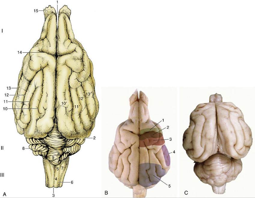
FIG. 8.23 (A) Dorsal view of the canine brain. I, Cerebral hemispheres; II, cerebellum; III, medulla oblongata. 1, Longitudinal fissure; 2, transverse fissure; 3, dorsal median sulcus; 4, tractus gracilis; 5, nucleus gracilis; 6, tractus cuneatus; 7, nucleus cuneatus; 8, cerebellar hemisphere; 9, cerebellar vermis;
10, marginal sulcus; 10', marginal gyrus; 11, ectomarginal sulcus; 11', ectomarginal gyrus; 12, suprasylvian sulcus; 12', suprasylvian gyrus; 13, ectosylvian sulcus; 13', ectosylvian gyrus; 14, cruciate sulcus; 15, olfactory bulb. (B) The real specimen of the dog. 1, motor cortex; 2, overlap of motor and somatosensory cortex; 3, somatosensory cortex; 4, auditory cortex; 5, visual cortex. (C) The real specimen of the cat.
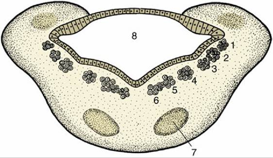
FIG. 8.24 Schematic transverse section of the metencephalon. The special somatic afferent nuclei are not shown. 1, Somatic afferent column; 2, visceral afferent column; 3, special visceral afferent column; 4, visceral efferent column; 5 and 6, somatic efferent column; 7, nuclei of pons; 8, fourth ventricle.
------- 1---------- Λ-------------------- 2---------------------- 3
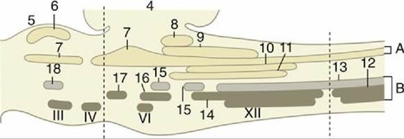
FIG. 8.25 Schematic representations of the brainstem showing the nuclei in an adult mammal. Roman numerals are used for nuclei of some cranial nerves. (A) Afferent nuclei; (B) efferent nuclei. 1, Mesencephalon; 2, rhombencephalon; 3, spinal cord; 4, cerebellum; 5, tectum mesencephali; 6, rostral colliculus (special somatic efferent [SSA]); 7, trigeminal nuclei (somatic efferent [SA]); 8, cochlear nuclei (SSA); 9, vestibular nuclei (SSA); 10, solitary nucleus of VII, IX, X (visceral afferent [VA]); 11, 11', gustatory nuclei of VII, IX (special visceral afferent [SVA]), respectively; 12, motor nucleus of XI (somatic efferent [SE]); 13, motor nucleus of X (visceral efferent [VE]); 14, nucleus ambiguus of IX, X (SE); 15, salivatory nuclei of VII, IX (VE); 16, motor nucleus of VII (SE); 17, motor nucleus of V (SE); 18, parasympathetic nucleus of III (VE).
» TABLE 8.2
Cranial Nerve Componentsa
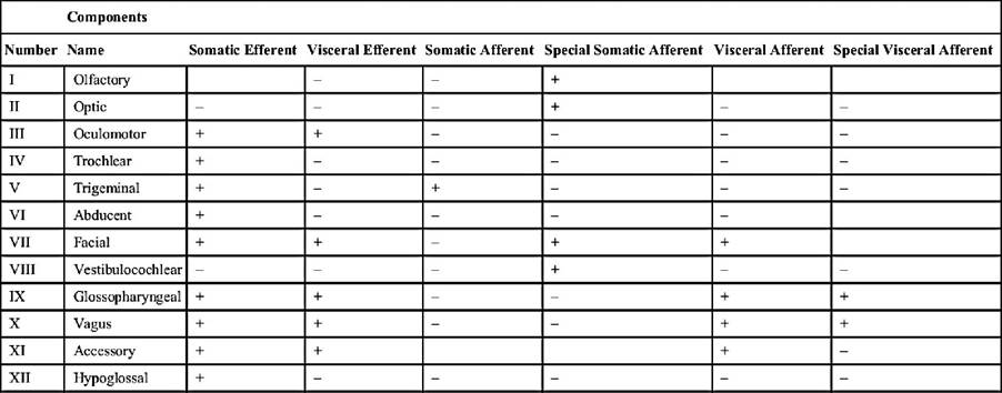
a Certain points are controversial: notably, the nerve trunks followed by fibers conveying proprioceptive information from various muscles of the head and the precise distribution of the medullary component of the accessory nerve.
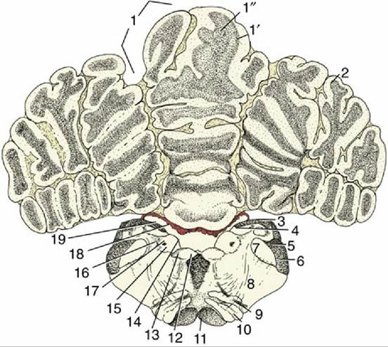
FIG. 8.26 Transverse section of the canine brain at the level of the hypoglossal nerve (XII). 1, Cerebellar vermis; 1', cortex; 1", medulla; 2, cerebellar hemisphere; 3, fasciculi gracilis and cuneatus; 4, gracile and cuneate nuclei; 5, caudal cerebellar peduncle; 6, spinal tract of the trigeminal nerve; 7, nucleus of the spinal tract of the trigeminal nerve; 8, reticular formation; 9, root of hypoglossal nerve; 10, caudal olivary nucleus; 11, pyramidal tract; 12, medial longitudinal tract; 13, motor nucleus of XII; 14, sulcus limitans; 15, motor nucleus of X; 16, solitary tract (special visceral afferents of VII, IX, and X); 17, solitary nucleus; 18, choroid plexus; 19, fourth ventricle.
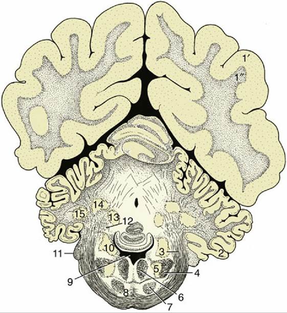
FIG. 8.27 Transverse section of the canine brain at the level of the middle cerebellar peduncle. 1', 1", cerebral hemisphere: 1', neocortex; 1", fibers; 2, paraflocculus lateralis; 3, middle cerebellar peduncle; 4, spinal tract of the trigeminal nerve; 5, nucleus of the spinal tract of the trigeminal nerve; 6, medial
longitudinal fasciculus; 7, pyramidal tract; 8, pontine nuclei; 9, fourth ventricle; 10, nuclei of the vestibulocochlear nerve (VIII); 11, root of VIII; 12, rostral cerebellar peduncle; 13, fastigial nucleus; 14, nucleus interpositus; 15, lateral cerebellar nucleus.
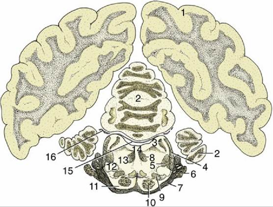
FIG. 8.28 Transverse section of the canine brain at the level of the trigeminal nerve. 1, Cerebral hemisphere; 2, cerebellum; 3, rostral cerebellar peduncle; 4, lateral lemniscus; 5, rubrospinal tract; 6, root of V; 7, middle cerebellar peduncle; 8, medial longitudinal fasciculus; 9, medial lemniscus; 10, pyramidal tract; 11, pontine nuclei; 12, nucleus of lateral lemniscus; 13, reticular formation; 14, fourth ventricle; 15, rostral medullary velum; 16, root of IV.
Axons traveling in the three cerebellar peduncles form the attachments of the cerebellum on each side. The cerebellum is also attached to the brainstem by the caudal and rostral medullary vela (Fig. 8.21/15,15'). The caudal peduncle (Fig. 8.22/10) connects the cerebellum with the medulla oblongata and is largely composed of afferent fibers entering the cerebellum, of which some run from origins within the spinal cord and others run from the vestibular nuclei, the olivary nucleus, and the reticular formation. The middle peduncle (brachium pontis; Fig. 8.22/9) is also composed of afferent fibers; these arise from pontine nuclei. The rostral peduncle (brachium conjunctivum; Fig. 8.22/11) is attached to the midbrain; it is largely composed of efferent fibers leaving the cerebellum to synapse in the red nucleus, reticular formation, and thalamus. The rostral peduncle also includes a considerable afferent component that continues the ventral spinocerebellar tract. The three peduncles are compressed closely together at their attachments to the cerebellum.
The functions of the cerebellum are concerned with the control of balance, the coordination of postural and locomotor activities, and motor planning. Balance, including head and eye movements, is largely controlled within the flocculonodular node. The vermis and medialmost portions of the hemispheres are concerned with the feedback regulation of motor function of axial and limb muscles. The lateral hemispheres are involved in planning motor movements. There is somatotopic representation in the cerebellar cortex, in that adjacent regions of the body are represented by corresponding adjacent areas in the cortex.

FIG. 8.29 Schematic transverse section of the mesencephalon. 1, Tectum; 2, tegmentum; 3, crus cerebri; 4, mesencephalic aqueduct; 5, oculomotor nucleus (III); 6, red nucleus; 7, substantia nigra; 8, locus coeruleus.
The cerebellum does not project directly to lower motor neurons and so does not initiate movement. Instead, the cerebellum coordinates movements by continually adjusting the output of upper motor neurons. Thus, damage to the cerebellum produces incoordination but not paralysis.