The Spinal Cord
The spinal cord (medulla spinalis) is an elongated structure that is more or less cylindrical but with some dorsoventral flattening and certain regional variations in form and dimensions.
The most important of these are the thickenings (intumescentiae; Fig. 8.15) of the regions that give origin to the nerves supplying the forelimbs and hindlimbs and the final caudal tapering (conus medullaris). The spinal cord lies within the vertebral canal, formed by the alignment of the series of vertebrae, and is divided into segments along its craniocaudal length. Each spinal segment is defined by its association with a pair of spinal nerves, formed by the union of a dorsal root arising from dorsolateral margin of the spinal cord, containing sensory afferent fibers, and a ventral root, containing motor efferent fibers, arising ventrolaterally as described (p. 27). The segments, and corresponding spinal nerves, are grouped and named according to the region of the body: from cranial to caudal, these are cervical, thoracic, lumbar, sacral, and caudal. The number of segments in each group varies among species and is identical to the corresponding number of vertebrae for each species except in the cervical region, where there is always one less cervical vertebra than cervical spinal segment. The position of the spinal segments relative to the corresponding vertebrae varies along the length of the cord. Species-specific details are discussed in later chapters, but the most consistent and notable disparity is in the lumbar region, where the short craniocaudal length of each of the caudalmost lumbar segments and of the sacral and caudal spinal segments results in the cranial displacement of these segments to lie within the lumbar vertebrae. The relationship between the spinal nerves and corresponding vertebrae remains consistent along the length of the spinal column, however, with the result that the last lumbar, and the sacral and caudal spinal nerves necessarily must travel caudally within the vertebral canal before exiting at their corresponding vertebrae. This group of nerves, thought to resemble the tail of a horse, is termed the cauda equina and represents the innervation of much of the perineum, tail, and pelvic viscera (Fig. 8.15/6 and Fig. 12.9/9). The location of the cauda equina serves as a convenient access for anesthesia of these structures, particularly in obstetric cases.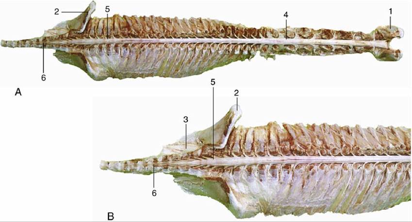
FIG. 8.15 (A) Dorsal view of the spinal cord and the vertebral pedicles of the horse. The spinal cord is shorter than the vertebral canal (ascensus medullae spinalis). (B) Enlargement of the caudal part. 1, Atlas;
2, ilium; 3, sacrum; 4, cervical intumescence; 5, lumbar intumescence; 6, cauda equina.
A simple transverse section shows a central mass of gray matter perforated in the midline by a small central canal, which is the derivative of the lumen of the embryonic neural tube (Figs. 8.13/1 and 8.11A and B/2). The gray matter, which has a crude resemblance to a butterfly or an H, is commonly described as exhibiting dorsal and ventral horns or columns; the former is a rather misleading term because the horns extend the length of the cord (Fig. 8.16). The dorsal horn corresponds to the embryonic alar plate. It contains neurons receiving afferent input from sensory axons entering the cord through the dorsal root as well as innumerable interneurons. Somatic sensory input synapses on dorsomedially located interneurons and visceral afferent neurons synapse on dorsolaterally located interneurons (Fig. 8.17). The ventral horn is derived from the basal plate; it is composed in part of the cell bodies of somatic efferent, or motor neurons, which are located ventrally, and visceral efferent neurons, which form an additional lateral horn confined to the thoracolumbar and sacral regions of the cord.
The neurons within each column are more specifically grouped according to their functional and topical associations, but this grouping is not grossly discernible.
The white matter that surrounds the gray matter is divided into three funiculi on each side (Fig. 8.18/I-III). The dorsal funiculus is contained between a shallow dorsal sulcus, extended deeply by a median glial septum, and the line of origin of the dorsal roots of the spinal nerves (see Fig. 8.13). The lateral funiculus is contained between the lines of the dorsal and ventral roots, and the ventral funiculus between the line of the ventral roots and a ventral fissure that penetrates far into the white matter, although it leaves a considerable commissure connecting the right and left halves. This ventral fissure is occupied by a mass of pia that appears as a glistening streak on the ventral surface of the cord.
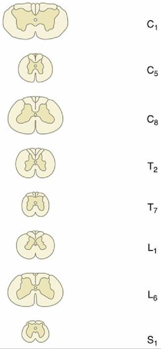
FIG. 8.16 Transverse sections of the canine spinal cord (the vertebral levels are indicated: C, cervical; T, thoracic; L, lumbar; S, sacral). Note the changes in diameter of the cord and in the relative proportions of gray (darker) and white (lighter) substance.
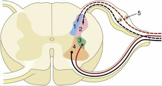
FIG. 8.17 Schematized subdivision of the gray substances in the spinal cord. 1, Input from somatic afferent neurons; 2, input from visceral afferent neurons (1 and 2 form the dorsal horn); 3, visceral efferent neurons; 4, somatic efferent neurons (3 and 4 form the ventral horn); 5, dorsal root ganglion.
The funiculi are composed of ascending and descending nerve fibers, of which many are grouped within bundles (fasciculi or tracts) of common origin, destination, and function (see Fig. 8.18). Although the details vary among species, the dorsal funiculus is almost entirely composed of ascending tracts (Fig. 8.18/1 and 2), as are the most lateral regions of the lateral funiculi (Fig. 8.18/5 and 6). Most of the lateral funiculi and the ventral funiculi contain both ascending and descending tracts.